If you have lower back pain with leg symptoms, there is a good chance your imaging report used the words "disc bulge" or "disc herniation." Most patients hear these terms, assume they mean more or less the same thing, and walk away confused about what they actually have and what to do about it. Clinicians sometimes use them interchangeably too, which adds to the frustration.
They are not the same. The distinction matters both for prognosis and for choosing the right conservative care path. Here is the complete breakdown.
What a disc is and what it does
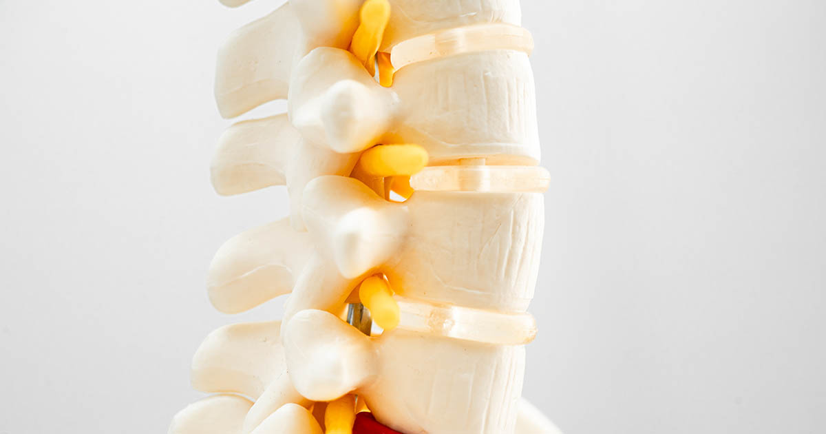
Each intervertebral disc sits between two vertebrae and acts as a shock absorber and spacer. The disc has two main layers. The outer ring is the annulus fibrosus: a tough, laminated collagen structure that contains the disc contents under load. The inner core is the nucleus pulposus: a gel-like material with high water content that distributes compressive forces evenly across the vertebral end plates.
In a healthy disc, the nucleus stays centered. The annulus holds firm. When you bend, twist, lift, or compress the spine, the disc deforms slightly and springs back. That cycling motion is part of how discs receive nutrition, since adult discs are avascular (they have no direct blood supply) and depend on this pumping action to draw in fluid and nutrients.
Problems begin when the annulus weakens and the nucleus shifts, pushes outward, or escapes entirely.
What a disc bulge actually means
A disc bulge occurs when the outer annulus weakens and the disc extends beyond its normal footprint, but the annular fibers are still intact. Think of a hamburger patty pressing out past the edge of the bun. The nucleus has not broken through the annulus. The disc is deforming symmetrically or asymmetrically, but the outer wall is still in one piece.
On MRI, a bulge typically shows as a smooth, broad-based disc contour extending beyond the vertebral body margin by more than 25 percent of the disc circumference but without a focal protrusion. Radiologists often describe it as a "diffuse bulge" or "broad-based bulge."
Key features of a disc bulge:
- The annulus is weakened but not torn through.
- The nucleus is under pressure but still contained.
- Symptoms, if any, tend to be local back pain rather than strong nerve-root pain.
- Bulges can be symptomatic or asymptomatic. Many adults over 40 have one or more bulging discs on MRI with no meaningful pain.
- Bulges can progress to protrusions or herniations under continued load or with acute trauma.
What a disc herniation actually means
A herniation is a displacement of disc material beyond the normal disc space. The annulus has been compromised in some way, and disc content has moved through or past it. Herniations are classified by how far that material has traveled and whether the annulus is still connected to it.
There are four stages of herniation, in order of severity:
- Protrusion. The nucleus has pushed asymmetrically against a weakened section of annulus, creating a focal bulge. The annular fibers are still intact (or nearly so), but the disc has a clear focal protrusion rather than a smooth broad-based bulge. On MRI: the disc extends outward at one focal point. This is sometimes still called a "herniation" even though the annulus technically has not fully ruptured.
- Extrusion. The nucleus has pushed through the annulus fibers. The herniated material is still connected to the disc by a "neck" of disc tissue. This is a true herniation in the classic sense and is the most commonly treated stage. Nerve root compression is common here. Symptoms often include leg pain (radiculopathy), tingling, and weakness.
- Sequestration (free fragment). The herniated material has separated completely from the disc and is now a free fragment inside the spinal canal. This is the most severe stage. The free fragment can migrate up or down the canal. Symptoms can be severe and may include bilateral leg symptoms or bladder or bowel changes, which are red flags requiring immediate evaluation.
- Resorption. Not a stage of worsening but of resolution. The body sometimes reabsorbs herniated disc material over months to years. Larger herniations paradoxically have a higher rate of spontaneous resorption than small ones, because the immune system recognizes the escaped nucleus as foreign and mounts an inflammatory response that gradually breaks it down.
Spontaneous resorption is real. Research has documented herniation reduction on follow-up MRI in patients who never had surgery. Conservative care supports that process by managing inflammation, maintaining circulation to the area, and keeping the nerve root decompressed enough to function while healing proceeds.
How the two cause different symptoms
The symptom difference between a bulge and a herniation is mostly a function of whether a nerve root is being mechanically compressed and irritated.
Disc bulge symptoms tend to be:
- Aching or stiffness in the low back.
- Pain that worsens with prolonged sitting or forward bending (loading the disc).
- Mild referred discomfort into the buttock or upper thigh, but not sharp shooting leg pain.
- Symptoms that respond relatively well to activity modification and conservative care.
Disc herniation symptoms tend to be:
- Sharp, shooting, or burning pain that travels down the leg, often into the calf or foot. This is the classic radiculopathy pattern.
- Tingling or numbness in a dermatomal (nerve-distribution) pattern. L4 herniation affects the inner calf. L5 affects the outer calf and top of foot. S1 affects the heel and outer foot.
- Weakness: difficulty raising the heel (S1), dropping the foot when walking (L4/L5).
- Pain that is worse with Valsalva maneuvers: coughing, sneezing, bearing down. Increased intradiscal pressure briefly worsens the nerve compression.
- Pain that is sometimes better with movement (walking) but worse after sitting or standing still for long periods.
Overlap exists. A large bulge can compress a nerve root just as effectively as a protrusion. A small protrusion near an already-inflamed nerve root can produce severe symptoms out of proportion to the structural finding on imaging. MRI findings and clinical symptoms are notoriously inconsistent, which is one reason the clinical exam is still the most important diagnostic step.
Why imaging alone does not tell the whole story
MRI reports frequently find disc bulges and herniations in adults who have no symptoms whatsoever. A landmark 2015 study in the American Journal of Neuroradiology imaged 3,110 asymptomatic adults and found disc degeneration in 37 percent of 20-year-olds, climbing to 96 percent of 80-year-olds. Disc bulges were present in 30 percent of asymptomatic 20-year-olds and 84 percent of 80-year-olds. Herniations were present in 29 percent of 20-year-olds with no back pain at all.
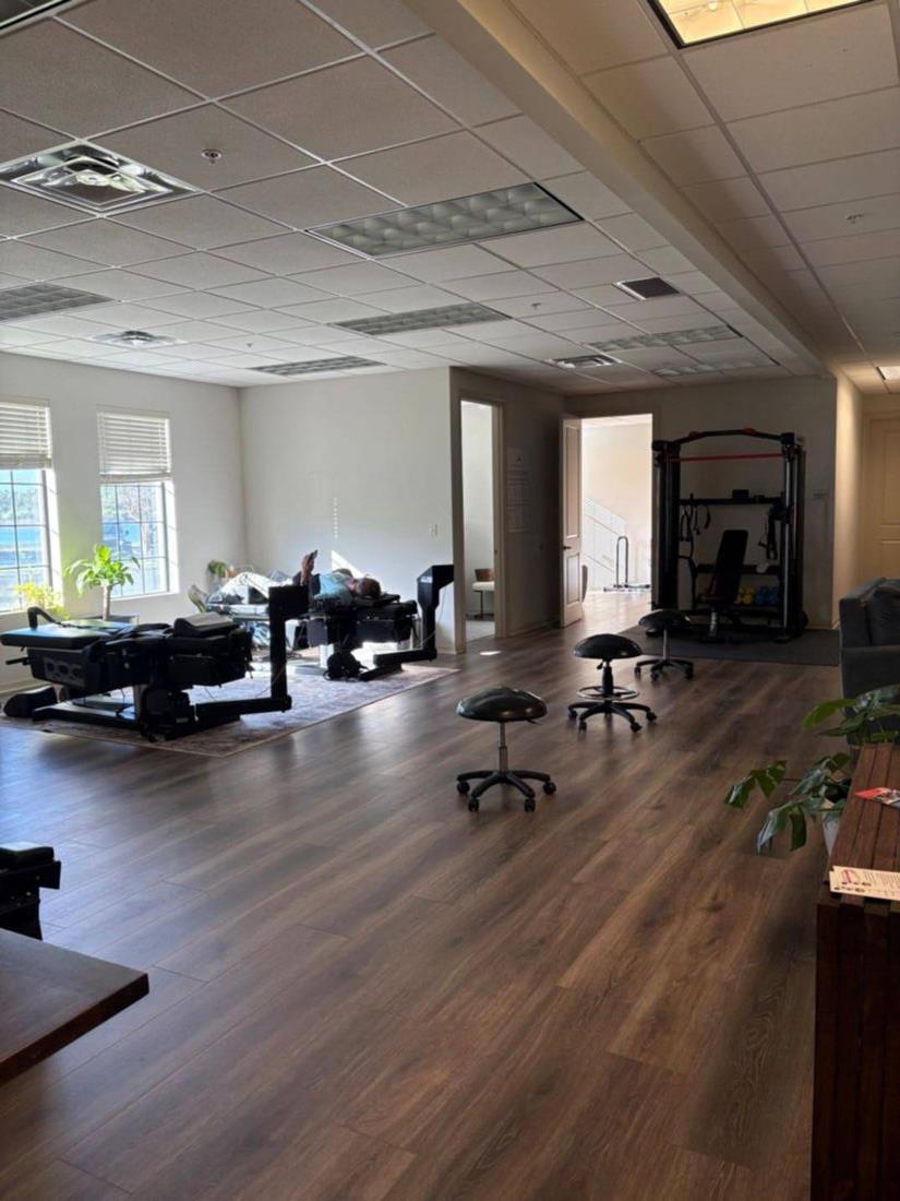
What this means clinically: the imaging finding does not tell you whether that disc is causing your pain. The exam does. A provider who reads your MRI without examining you is providing an incomplete picture. At our Lakewood Ranch office, Dr. Banman reviews imaging alongside orthopedic and neurological testing, range of motion, and symptom provocation testing. The goal is to correlate what the images show with what the body is actually doing.
Treatment paths: where bulge vs herniation matters
For conservative care, the bulge versus herniation distinction influences the aggressiveness and type of treatment rather than the basic approach. Both conditions respond well to conservative care in the majority of cases.
For disc bulges without nerve root involvement:
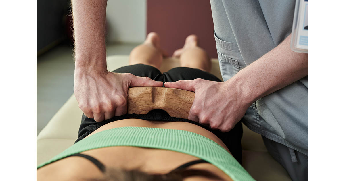
- Spinal decompression to reduce intradiscal pressure and restore disc hydration.
- Spinal manipulation or mobilization where appropriate.
- Core stabilization and postural retraining to reduce load on the affected segment.
- Activity modification: reducing prolonged forward flexion and seated load.
For disc herniations with nerve root involvement:
- Spinal decompression is often the first-line mechanical approach. Computer-guided traction creates negative intradiscal pressure that can draw herniated material back toward center and reduce direct mechanical pressure on the nerve root. The protocol is more structured than for bulges: controlled angles, precise tension curves, and a progressive session count.
- Class IV laser therapy to reduce nerve inflammation and support tissue healing around the compressed root.
- EMS (electrical muscle stimulation) to maintain muscle function in inhibited muscle groups during recovery.
- Careful adjustment protocols that do not load the herniated segment into further protrusion.
For severe herniations with progressive neurological deficits, surgical consultation is appropriate. Red flags that change the calculus: rapidly progressing weakness, loss of bowel or bladder control, or saddle anesthesia (numbness in the groin and inner thighs). Any of these warrants an immediate ER visit rather than a chiropractic evaluation.
For the large middle group of herniations with nerve pain but intact neurological function, conservative care with a structured decompression protocol gives many patients a real shot at avoiding surgery. Some are not surgical candidates anyway due to age or co-morbidities, making conservative care the only realistic option. Outcome data on non-surgical management of lumbar disc herniation shows that 60-90 percent of patients with non-progressive radiculopathy improve meaningfully with conservative care over 3-6 months.
The disc spectrum: it is a continuum, not categories
One of the most useful ways to think about disc problems is as a continuum rather than discrete categories. Healthy disc, early degeneration, disc desiccation (loss of water content), annular fissures, focal protrusion, extrusion, sequestration. Most patients arrive somewhere in the middle of that spectrum, and many have findings at multiple levels.
Treatment decisions are made based on where someone is on that spectrum and what direction they are heading. A contained bulge in a 35-year-old that has been stable for two years is a different problem than a bulge that became a protrusion over three months in a 55-year-old with new leg symptoms. The imaging category is just one data point.
What to ask your provider
If you have received an MRI report describing a bulge or herniation, here are the questions worth asking at your next appointment:
- Is the finding at this level actually correlating with my symptoms (dermatomal pattern, provocation testing)?
- Is there any progressive neurological deficit that changes the urgency of this?
- Is this a contained vs. non-contained herniation, and does that affect care options?
- Am I a candidate for spinal decompression?
- What does the disc height at this level look like relative to the levels above and below (loss of disc height limits decompression traction effectiveness)?
- Is there facet joint involvement at the same level that might require its own treatment?
These questions help you move from "I have a herniated disc" (a structural label) to "here is what is specifically driving my symptoms and what the best conservative care path looks like for this presentation" (a working clinical plan).
If you are in the Lakewood Ranch, Bradenton, or Sarasota area and want your imaging reviewed in context of a full clinical exam, Dr. Banman offers comprehensive evaluations that include orthopedic testing, range of motion assessment, neurological screening, and imaging correlation. See our conditions page for a full list of what we evaluate and treat, or visit our spinal decompression page for more on that specific intervention.