Patients hear "spinal decompression" and picture one of two things: either a surgeon cutting something out, or a simple stretching device they could find at a sporting goods store. Neither is accurate. The computer-controlled decompression table we use in our Lakewood Ranch office is a specific therapeutic tool with a specific mechanism, and understanding that mechanism is the first step to knowing whether it belongs in your care plan.
This post covers the actual physics, the conditions where decompression tends to produce the best results, the conditions where it probably will not help much, and the things patients often expect from it that no amount of traction will deliver.
The Basic Mechanism: Negative Intradiscal Pressure
Your intervertebral discs do not have a direct blood supply. They rely on a process called imbibition: when pressure inside the disc drops, the disc draws in water, oxygen, and nutrients from the surrounding tissue. When pressure stays chronically elevated, that flow reverses. The disc gradually dries out and starves.
Spinal decompression works by creating a brief, controlled reduction in intradiscal pressure, measured in research settings as negative pressure (below atmospheric). In a healthy disc, everyday movement handles this naturally. In a disc that is herniated, dried out, or sitting under the prolonged load of desk work or poor posture, that self-regulating cycle breaks down.
A computerized decompression table applies a precise pulling force to your lumbar or cervical spine in a way that resists the muscle guarding reflex. Manual traction, inversion tables, and hanging from a pull-up bar all create a stretch, but the muscles respond by tightening to protect the spine, which counteracts the force. The decompression table varies the pull in a logarithmic ramp-up and ramp-down pattern that stays below the threshold that triggers that guarding response. That is the key engineering difference between a DOC decompression table and a basic traction unit.
The goal is not to forcefully pull the spine apart. The goal is to unload the disc below the point where muscles grab back, so the disc itself can respond to the change in pressure.
What Happens to a Herniated Disc During Decompression
When intradiscal pressure drops below a critical threshold, two things tend to happen. First, the posterior annular fibers (the tough outer ring of the disc) have more space to relax. Second, the negative pressure acts like a gentle vacuum on the nucleus pulposus (the soft inner material that herniates through the annular tears).
Research using real-time MRI during decompression has documented measurable changes in disc geometry during sessions. These are not permanent single-session transformations. What researchers and clinicians observe is a cumulative effect over a series of sessions: the herniation may retract partially, annular fibers may partially re-approximate, and the disc height may increase slightly as hydration improves.
Our spinal decompression program is structured around this cumulative effect. A single session produces a transient change in pressure. A full course of 15 to 20 sessions over 6 to 8 weeks works to create conditions where those changes can consolidate.
This also explains why patients sometimes feel worse in the first few sessions. As the disc rehydrates and begins to shift position, it can temporarily press more firmly against nerve tissue before it starts pulling back. Most patients who persist past the 4th or 5th session report a turning point where the pattern reverses.
Who Tends to Respond Well
Based on what we see clinically, the patients who tend to have the strongest responses to decompression share a few characteristics.
Disc herniation with nerve root symptoms
When a herniated disc at L4-L5 or L5-S1 is pressing on the sciatic nerve and producing leg pain, foot tingling, or weakness, decompression directly addresses the mechanical cause. The retraction potential (whether the herniation can be pulled back) is best in patients who still have relatively good disc height. A disc that has almost completely collapsed has less structural material to work with.
For more on this pattern, see our post on disc-driven sciatica.
Degenerative disc disease with preserved disc height
Patients with moderate disc degeneration who still have measurable disc height on imaging tend to respond better than those with severe end-stage degeneration. The disc needs something to decompress. When the nucleus has fully desiccated and the vertebral bodies are close to bone-on-bone contact, the physics of the decompression mechanism change significantly.
Lumbar stenosis in the early to moderate stage
Spinal stenosis creates nerve pressure through a different mechanism (narrowed canal rather than herniated material pressing outward), but decompression can still reduce the overall mechanical load on the area. Many stenosis patients report that the flexion-distraction component of the treatment gives them relief that holds longer than manual therapy alone. This is the pattern described in our post on why stenosis worsens with walking.
Failed conservative care that has not yet reached surgical threshold
Many patients we see have tried physical therapy, medication, or basic chiropractic adjustments without lasting improvement. If imaging shows disc pathology as the underlying cause, decompression is often the next logical step before a surgical consultation. It addresses the disc directly in a way that massage, stretching, and adjustment alone cannot replicate.
What Decompression Does Not Do
This section matters as much as the above. A therapy that is genuinely useful in some situations is not useful in all situations, and the marketing around decompression equipment has unfortunately blurred those lines.
It does not fix facet joint arthritis
Facet joints are the small paired joints at the back of each vertebral segment. When these degenerate, the pain pattern is often stiffness-driven, worse in the morning, and aggravated by extension (leaning backward). Decompression creates primarily an axial unloading force. It reduces disc pressure but does not directly address the facet joint surfaces. Facet-dominant pain often responds better to mobilization, manipulation, and targeted muscle strengthening than to decompression.
It does not resolve severe nerve damage
If the sciatic nerve or a cervical nerve root has been compressed for a long time, the nerve itself may have suffered structural damage. Decompression removes the mechanical source of compression and gives the nerve the best possible environment to recover. But it does not regenerate axons or repair myelin. That process, when it happens, takes months and depends on the extent of the original injury.
For patients with significant nerve damage, decompression is typically paired with Class IV laser therapy, which has evidence for accelerating peripheral nerve recovery through photobiomodulation.
It does not work well with certain structural instabilities
Spondylolisthesis (one vertebra sliding forward on the one below it) is a contraindication in moderate to severe grades. Pulling on an already-unstable segment risks making the slip worse. Patients with grade 2 or higher spondylolisthesis on imaging are not candidates for standard decompression, and any clinic offering it without reviewing your imaging is not doing the proper intake evaluation.
It is not a substitute for surgical intervention in the right cases
Certain presentations require surgery: cauda equina syndrome (loss of bowel or bladder control from disc pressure), progressive motor weakness, or spinal cord compression in the cervical spine. These are neurological emergencies. Decompression is a conservative tool. If those red flags are present, the referral goes to a spine surgeon, not to the decompression table.
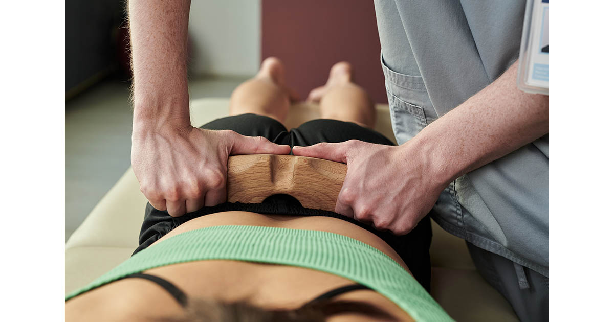
The Treatment Protocol: What to Actually Expect
A standard lumbar decompression session at our office runs 20 to 30 minutes on the table. The patient is fitted with a pelvic harness that anchors to the lower section of the DOC table. The upper body is stabilized. The computer program applies the prescribed force in the logarithmic pattern described above, cycling between pull and partial release throughout the session.
Before the first session, we review imaging (X-ray, MRI if available) to confirm the disc levels involved and rule out contraindications. The starting force is conservative, typically 40 to 50 percent of body weight for lumbar, and we build incrementally based on response.
Most patients describe the sensation as a gentle stretching or pulling with no sharp pain. If the pressure is uncomfortable or produces a new sharp symptom, that is feedback we adjust to immediately. The goal at every session is to stay in the therapeutic window, not to push through discomfort.
- Sessions 1 to 5: Establishing tissue tolerance, often modest immediate relief followed by some post-session soreness
- Sessions 6 to 10: Most patients report a turning point where leg pain begins to centralize (move from the foot back toward the spine), which is a sign the herniation is responding
- Sessions 11 to 20: Consolidation phase, extending the duration of relief between sessions and adding rehabilitative exercise to stabilize the disc
We typically pair decompression with whole body vibration as part of the post-session protocol. The vibration activates deep spinal stabilizers that hold the disc in its improved position after the session. See our post on why decompression and whole body vibration work better together for the full rationale.
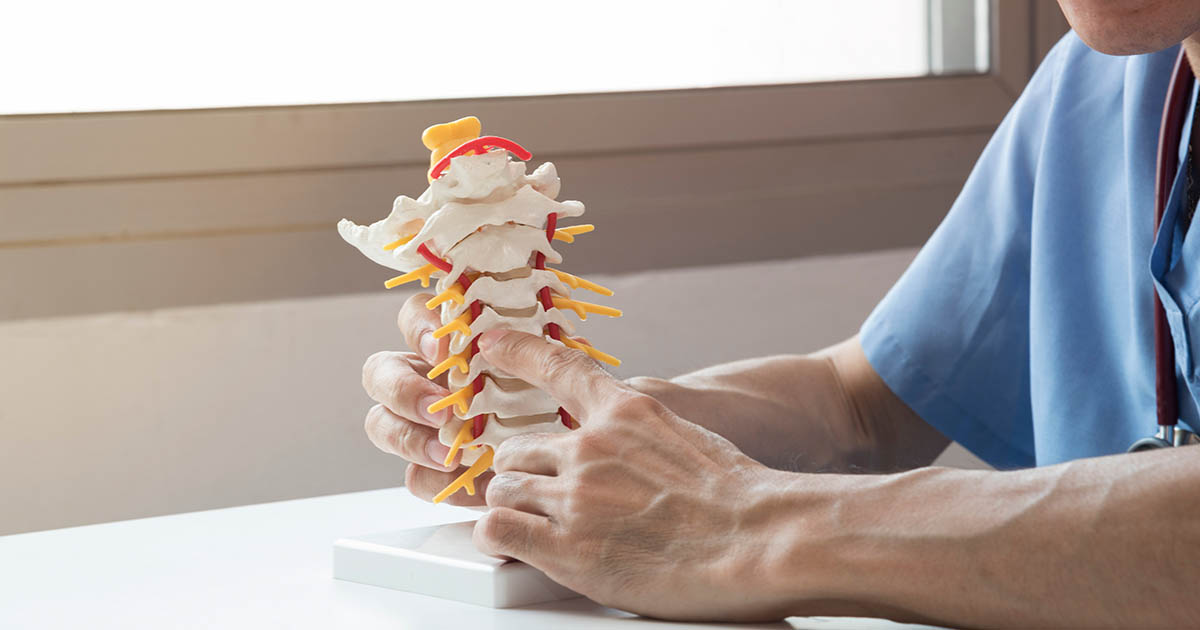
Cervical Decompression: The Same Physics, Different Setup
The same negative-pressure principle applies to the cervical spine (neck), and we offer cervical decompression for disc herniations pressing on the C5, C6, or C7 nerve roots. The setup is different: the patient is supine (face up), and the harness fits around the skull rather than the pelvis. Force levels are much lower, typically 10 to 20 pounds at peak, because the cervical spine structures are significantly smaller.
Cervical decompression tends to help patients who have arm pain, hand tingling, or weakness that originates from a confirmed cervical disc herniation. It is not the right tool for tension-type headaches, migraines, or neck pain that is purely muscular. A thorough evaluation determines which cases fit.
Combining Decompression With Other Therapies
In our experience, decompression produces the most durable results when it is part of a broader plan rather than the only intervention. Here is how it fits with the other tools we use:
- Class IV laser: Reduces inflammation in the surrounding soft tissue and nerve roots, which makes the disc more responsive to decompression and accelerates nerve recovery
- Electrical muscle stimulation (EMS): Activates inhibited paraspinal muscles that have shut down around the injured disc, improving spinal support once the disc begins to recover
- Chiropractic adjustment: Restores segmental motion at levels adjacent to the decompressed disc, reducing the compensatory loading patterns that originally contributed to disc stress
- Rehabilitative exercise: The final phase that ensures the gains made on the table carry over into daily activity
The combination is deliberate. Decompression creates the opportunity. The supporting therapies help the body use that opportunity. Doing decompression in isolation, without addressing the muscular and movement patterns that loaded the disc in the first place, tends to produce results that do not hold as well long-term.
How to Know If You Are a Candidate
The most reliable way is a proper evaluation: a history, physical exam, orthopedic and neurological testing, and a review of any available imaging. The intake exam tells us which disc levels are involved, whether nerve involvement is present, what the disc height looks like, and whether any contraindications exist.
Some questions that can help you self-screen before calling:
- Do you have imaging (MRI or X-ray) showing a disc herniation, bulge, or degenerative disc disease?
- Is the pain worsening with sitting or with rest, rather than improving?
- Do you have leg pain, foot tingling, or weakness that seems to come from the back rather than from the leg itself?
- Have you tried physical therapy or standard chiropractic adjustments for at least 6 to 8 weeks without meaningful improvement?
If most of those are yes, decompression is worth a conversation. If none of those fit, a different diagnostic pathway probably makes more sense first.
For patients wondering why rest seems to make their back pain worse rather than better, that pattern is a strong indicator of disc involvement. See our post on why back pain worsens after rest for more on the disc imbibition mechanism behind that symptom.