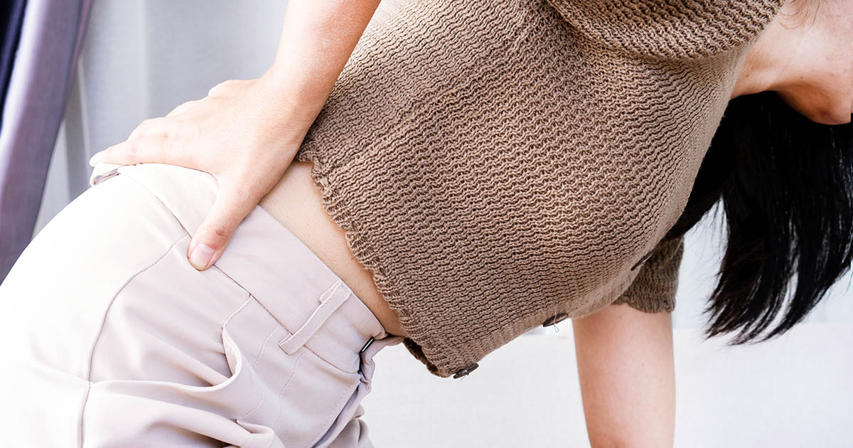
A lot of people in Lakewood Ranch describe the same scenario. They head out for a walk, feel reasonably okay for the first five or ten minutes, then notice a dull ache building in the lower back. It spreads into the buttocks, sometimes into one or both legs. By fifteen or twenty minutes in, they have to stop and rest. They sit down on a bench or lean forward on a cart at the grocery store, and within a few minutes the pain backs off enough to keep going.
This pattern has a name: neurogenic claudication. It is the hallmark symptom of lumbar spinal stenosis. And it is one of the more reliably diagnosable patterns in spinal medicine because the mechanics behind it are so specific.
If this sounds familiar, you are not imagining it, and it is not simply a matter of being out of shape. The canal that houses your spinal cord and nerve roots is narrowed, and walking loads that canal in a way that sitting and leaning forward do not. Understanding why that happens is the first step toward doing something about it.
What Is Happening Inside a Stenotic Spine
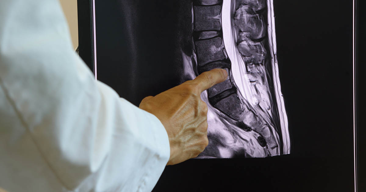
The spinal canal is a bony tunnel that runs through the center of your vertebral column from the base of your skull down to the sacrum. It houses the spinal cord in the upper and mid-spine, and a bundle of nerve roots called the cauda equina in the lower lumbar region. The canal has a finite diameter. When structures inside or around it enlarge or shift inward, the available space for neural tissue shrinks.
In lumbar spinal stenosis, that narrowing most commonly happens through one or more of the following: arthritic bone spurs growing from the facet joints or the posterior margins of the vertebral bodies; thickening of the ligamentum flavum (the yellow ligament running along the back of the canal); bulging or herniated disc material pressing forward from the front; or forward slippage of one vertebral body over the one below it, a condition called spondylolisthesis.
Any of these changes individually may be tolerated. Most adults over 55 have some degree of lumbar degeneration that does not produce symptoms. The problem arises when the cumulative narrowing crosses a threshold where the nerve roots running through the canal are compressed under load, specifically the load that comes with walking upright.
Why Walking Specifically Makes It Worse
The answer comes down to spinal position and how it affects canal diameter. When you stand upright and walk, your lumbar spine moves into relative extension. The lower back has a natural inward curve (lordosis), and walking accentuates that curve. Extension, even the mild extension of normal standing posture, does two things to a stenotic canal.
First, the ligamentum flavum, which runs along the back wall of the canal, buckles slightly inward when the spine extends. In a healthy canal this does not matter. In a canal that is already narrowed, even a few millimeters of additional intrusion from the ligament can meaningfully compress neural tissue.
Second, the foraminal openings (the side channels where individual nerve roots exit the canal) narrow in extension and widen in flexion. Walking in an upright posture continuously loads those foramina in their narrowed state.
The result is that nerve roots carrying blood vessels and sensory fibers begin to experience reduced circulation and increasing pressure the longer you walk. This is what produces the burning, cramping, or heaviness in the legs. It mimics the sensation of vascular claudication (poor blood flow to the legs), which is why the two are sometimes confused. The key distinguishing factor: vascular claudication improves when you simply stop walking and stand still. Neurogenic claudication from stenosis requires you to sit or flex forward to get relief, because that is what opens the canal again.
The classic test: a patient who can walk the same distance uphill more comfortably than on flat ground almost certainly has lumbar stenosis. Walking uphill forces slight forward lean, which flexes the lumbar spine and opens the canal. Downhill walking, which increases lumbar extension, is typically worse.
The Shopping Cart Sign: A Pattern Worth Knowing
Clinicians call it the "shopping cart sign." Patients with lumbar stenosis can often push a grocery cart for considerably longer than they can walk unassisted, because leaning on the cart keeps the lumbar spine in slight flexion, partially decompressing the canal.
Other versions of the same pattern include:
- Being able to ride a stationary or regular bicycle without symptoms (cycling keeps the hips flexed and the lumbar spine neutral to slightly flexed).
- Finding it easier to walk up stairs than down them.
- Leaning forward on a counter or sink to relieve leg heaviness.
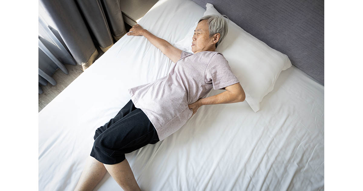
- Sleeping on your side with knees pulled up, finding that position relieves back and leg discomfort that lying flat aggravates.
- Sitting causing rapid relief of leg symptoms, while lying flat on your back may not.
If two or more of these describe your daily experience, that is a meaningful clinical signal. It does not confirm stenosis on its own, imaging is needed for that, but it points strongly in that direction.
Stenosis vs Disc Herniation: Why the Distinction Matters
Both conditions can produce radiating leg pain and back pain, and imaging will often show both in the same patient. But the clinical pattern differs in important ways, and treatment approaches differ accordingly.
Disc herniation typically involves a more acute onset, often tied to a specific movement or event. The leg pain from a herniation tends to follow a specific dermatomal pattern (a narrow band from the buttock down one leg, often to the foot), and it is frequently worse with sitting and forward flexion, not with walking. A patient with a purely herniated disc may actually feel better walking than sitting.
Stenosis, by contrast, tends to develop gradually over years and worsens with extended walking. The leg symptoms are often more diffuse and bilateral (both legs, though one may be worse). Rest relieves it reliably, but only if the rest involves spinal flexion.
This matters for care planning. Spinal decompression therapy, for example, uses distraction forces that put the lumbar spine into slight flexion and traction simultaneously, which addresses both the disc component and the canal pressure of stenosis. But the setup, angles, and duration differ based on which pattern is dominant. Treating a stenosis presentation like a pure disc herniation, or vice versa, tends to produce slower results. See our lumbar spinal stenosis page for more detail on how we approach the distinction clinically.
What Actually Causes Lumbar Stenosis to Develop
Stenosis is primarily a degenerative condition. It does not usually develop from a single injury or sudden event. It accumulates over years as the spine ages and the structures around the canal undergo normal degenerative changes.
The main drivers include:
- Facet joint arthritis: The facet joints at the back of each vertebral level are synovial joints, the same type as the hip and knee. As they degenerate, they develop osteophytes (bone spurs) that can grow into the canal or foramina.
- Ligamentum flavum hypertrophy: Chronic mechanical loading and inflammation cause this ligament to thicken. It runs along the posterior wall of the canal, so its thickening directly encroaches on neural space.
- Disc desiccation and collapse: As discs lose hydration and height with age, the foraminal height decreases, compressing nerve roots at their exit points. Degenerative disc disease and stenosis frequently coexist.
- Spondylolisthesis: When one vertebral body slips forward over the one below (a common finding at L4-L5 in older adults), it creates a step-off that narrows the central canal at that level.
- Congenital narrowing: A small percentage of patients are born with a naturally narrow canal. These individuals may develop symptomatic stenosis earlier and with less degeneration than average.
Florida's older demographic means stenosis is common in Lakewood Ranch, Bradenton, and Sarasota. It is one of the most frequent reasons adults over 60 seek evaluation at our office, and in many cases patients have been managing symptoms for years before getting a clear explanation of the mechanism.
What Conservative Care for Stenosis Looks Like
Surgery for spinal stenosis, specifically a laminectomy or laminotomy (removing bone to widen the canal), is sometimes appropriate. But the research on conservative versus surgical outcomes for lumbar stenosis is more nuanced than many patients realize, and a structured conservative program is appropriate as a first line for most presentations.
The goal of conservative care is to reduce the load on compromised neural tissue, restore as much available canal space as possible through positional and mechanical means, reduce inflammation in the affected structures, and improve the supporting musculature to reduce the compressive forces the canal experiences under load.
At Spine and Wellness Center Lakewood Ranch, a stenosis care plan typically involves several modalities working together:
Spinal decompression therapy uses a computer-controlled traction table that creates targeted distraction forces in the lumbar spine, placing the canal in a flexed and decompressed position for cycles of pull and release. Many patients with lumbar stenosis report progressive improvement in walking tolerance over the course of a decompression program. For more on how decompression works mechanically, visit our spinal decompression page.
Chiropractic care for stenosis focuses less on high-velocity manipulation (which is often modified or avoided in advanced stenosis) and more on restoring segmental mobility in the thoracic and lumbar spine, reducing the compensatory muscle patterns that develop when patients guard a painful area, and addressing the hip flexor tightness and quadratus lumborum shortening that accentuates lumbar extension.
Class IV laser therapy can reduce inflammation in the facet joints and surrounding soft tissue. This does not change the bony architecture of the canal, but reducing local inflammation reduces the edema that contributes to symptom severity, particularly in the ligamentum flavum and nerve root sleeves.
Whole-body vibration activates deep spinal stabilizers (multifidus and transversus abdominis) that are often inhibited in chronic back pain patients. These muscles are critical to reducing the compressive load on the canal during upright activity. Patients who stabilize their lumbar spine better tend to tolerate more walking before symptom onset.
Hyperbaric oxygen therapy is used in more severe or chronic nerve compression cases. Compressed nerve roots have reduced oxygen delivery; HBOT increases dissolved oxygen in the plasma, which can reach hypoxic tissue more effectively than hemoglobin-bound oxygen. We use it selectively for patients with significant neurological involvement or chronic refractory symptoms.
None of these modalities, alone or together, will reverse the bony narrowing visible on an MRI. What a structured program can do is reduce the symptom load to a manageable level, extend comfortable walking distance, and in many cases eliminate the need for surgical consultation.
When Surgery Does Come Up
There are situations where surgical decompression is the appropriate next step. Dr. Banman will tell you directly if your presentation suggests that conservative care is unlikely to be sufficient. The clearest indicators include:
- Progressive neurological deficit: Worsening weakness in a specific muscle group, loss of reflexes, or new bladder or bowel symptoms (the last is a medical emergency requiring immediate evaluation).
- Failure of a genuine structured conservative program: Not a few visits, but a complete, multi-modality effort over 8 to 12 weeks with no meaningful improvement in walking tolerance or pain levels.
- Imaging showing severe canal compromise: MRI findings of cord or cauda equina signal change indicating neural injury, rather than simply mechanical compression.
- Quality of life that is unacceptable even with modifications: Some patients with stenosis cannot manage daily function even with conservative measures, and the surgical benefit clearly outweighs the risk for their specific anatomy and health profile.
When surgical referral is appropriate, we coordinate with spine surgeons in the Sarasota and Tampa areas and can provide the clinical documentation from our examination and treatment course to assist in that evaluation.
For more on the range of back pain conditions we see and treat at our Lakewood Ranch office, and how they overlap with stenosis, that page gives a broader overview. For the nerve-related leg symptoms that often accompany stenosis, see our sciatica page for how we approach that distinction diagnostically.