At some point in their 40s, 50s, or 60s, millions of adults get an MRI for back pain and leave with a report containing the phrase "degenerative disc disease." Some receive this finding after years of chronic pain. Others get it almost incidentally, while the image was taken for something else entirely. Either way, the word "disease" tends to land hard.
In our Lakewood Ranch office, we see patients every week who came in genuinely scared because their MRI report made it sound like their spines are failing. Most of the time, the reality is far less alarming than the terminology suggests. In some cases, the finding is barely relevant to why they hurt at all. In other cases, the degeneration is real, active, and something we can actually address.
Understanding the difference matters. It changes what treatment makes sense, what to expect over the next year, and whether surgery is even a remote possibility.
What "Degenerative Disc Disease" Actually Means
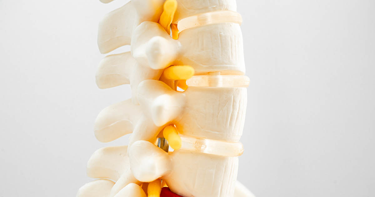
The name is a medical legacy problem. "Degenerative disc disease" (DDD) is not a disease in the way that diabetes or cancer is a disease. It is a descriptive term for age-related changes in the intervertebral discs: the rubbery, shock-absorbing structures that sit between each vertebra in your spine.
Each disc has two layers. The outer layer, called the annulus fibrosus, is a tough ring of fibrocartilage. The inner layer, the nucleus pulposus, is a gel-like core that is mostly water in younger people. That water content is what gives discs their ability to compress and rebound under load.
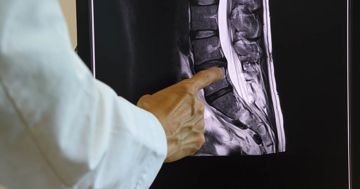
Over time, and especially after repeated mechanical stress, those discs lose hydration. The nucleus becomes drier and less pliable. The disc loses some height. The annulus may develop micro-tears. On an MRI, these changes show up clearly: darker signal on T2-weighted images (less water), narrowed disc spaces, and sometimes evidence of altered loading patterns in the adjacent vertebrae.
Radiologists and orthopedic specialists have standardized language for grading this process, typically from Grade I (normal, youthful disc) through Grade V (severe collapse and black disc appearance). Most people who receive a DDD diagnosis fall somewhere in the Grade III range: visible degeneration, meaningful disc height reduction, but not collapse.
The critical point: this is a spectrum. And where someone falls on that spectrum does not directly predict how much pain they have.
The Biology Behind Disc Aging
Discs age for several reasons, and not all of them are within your control. Genetic factors play a significant role. Studies of identical twins show that disc degeneration patterns often match between siblings even when their lifestyles differ substantially. Your disc biology is partly inherited.
Beyond genetics, the discs are unusual tissues: by adulthood, they have essentially no direct blood supply. They rely on a process called diffusion to bring nutrients in and waste products out, pumped through the endplates of the adjacent vertebrae. That diffusion is most efficient when the disc is loaded and unloaded cyclically, which is why normal movement actually helps disc health.
Prolonged static loading, particularly prolonged sitting, disrupts this diffusion cycle. So does smoking, which impairs circulation to the endplates. So does excess body weight, which increases baseline disc pressure. And so does repetitive heavy lifting with poor mechanics, which accelerates the annular micro-tear process.
In Florida's sedentary, car-dependent, heat-avoidance culture, many of the conditions for accelerated disc aging are present. Long drives on I-75, desk work, and reduced outdoor activity during the summer months all reduce the movement that keeps discs healthy. This does not mean DDD is inevitable for everyone, but it helps explain why the Lakewood Ranch population trends toward it.
Why DDD on Your MRI Does Not Always Mean Pain
This is the most important thing to understand about degenerative disc disease: the imaging finding and the pain experience are not the same thing.
Multiple large studies have scanned the lumbar spines of people with no back pain at all and found degenerative changes in a substantial portion of them. The numbers go up with age. By age 50, roughly 60 to 80 percent of people have some imaging evidence of disc degeneration. By 70, the rate approaches 90 percent. Most of these people are not in chronic pain. Many never had significant back pain in their lives.
A 2015 systematic review published in American Journal of Neuroradiology found that disc degeneration was present in 37 percent of asymptomatic 20-year-olds and 96 percent of asymptomatic 80-year-olds. The finding is extremely common. The symptoms are not inevitable.
What this means practically: if your radiologist found DDD and your back hurts, DDD may or may not be the cause of that pain. It could be the discs themselves. It could be secondary structures that are reacting to the disc changes. Or the pain could be coming from something else entirely, with DDD just coincidentally present on the image.
Good diagnostic work distinguishes between these possibilities. Symptom patterns, physical exam findings, and responses to specific provocative tests tell us far more about the actual pain generator than the imaging alone.
When DDD Becomes Symptomatic: The Patterns That Matter
When degenerative disc disease is genuinely driving pain, the symptom patterns tend to be recognizable. Understanding them helps you describe what you are experiencing accurately, which in turn helps us identify the right treatment approach.
Discogenic Pain
Pain that comes directly from the disc itself is called discogenic pain. Discs do have nerve endings in their outer layers (the annulus), and a degenerated disc with annular tears can generate real, sometimes severe, pain.
Discogenic pain is typically axial: it stays in the low back or mid-back, without traveling far down the leg. It is often described as a deep, dull aching, with sharp spikes during certain movements. It tends to be worst with flexion-based activities: bending forward, sitting for long periods, riding in a car. Many patients notice it spikes when transitioning from sitting to standing, then eases slightly as they walk around. The grab-and-groan moment when rising from a chair is a classic discogenic pattern.
Morning stiffness is another hallmark. Discs absorb fluid overnight while you are lying down (because the load is reduced), then have to adjust to the compressive forces of standing up. A degenerated disc handles this adjustment less smoothly. Back pain that is worse after rest and better with movement, but then worsens again with prolonged activity, is a pattern we see consistently with discogenic involvement.
Referred Pain
Degenerated discs can also produce referred pain: a vague, achy discomfort that spreads into the buttocks, hips, or even upper thighs without following a clear nerve root pattern. This is different from sciatica (which involves actual nerve compression and typically produces sharper, more radiating pain). Referred discogenic pain is often described as a dull spread, harder to localize precisely, and tends to shift rather than staying in one consistent location.
Secondary Structure Pain
When a disc degenerates and loses height, the mechanical relationships between adjacent structures change. The facet joints at the back of each vertebral level take on more load than they were designed to handle. They can develop their own inflammation and arthritis. The ligaments become looser. The muscles that support the segment often go into chronic guarding. These secondary sources of pain are common and often explain why a patient's symptoms feel more diffuse than a simple disc problem would suggest.
The Downstream Consequences of Long-Term DDD
Degenerative disc disease does not stay static in most people. Over years and decades, it can progress and generate secondary problems that are distinct from the DDD itself.
- Disc bulge and herniation. A degenerated disc with a compromised annulus is more susceptible to bulging outward or herniating (rupturing through the annular wall). When that herniated material contacts a nerve root, it produces radiculopathy: the sharp, electric, radiating pain into the leg or arm that most people call sciatica. This is a different problem from DDD itself, though it often develops in the context of DDD. See our post on disc herniation versus disc bulge for a full breakdown of the difference.
- Bone spurs (osteophytes). As the disc loses height and the vertebral endplates are exposed to altered loading, the bone responds by laying down extra structure. These bone spurs can sometimes encroach on nerve pathways and produce their own compression symptoms.
- Spinal stenosis. When DDD is combined with facet joint arthritis and ligament thickening over years, the overall space available for the spinal cord and nerve roots can narrow significantly. This produces lumbar spinal stenosis, a condition where pain builds with walking and eases when you sit or lean forward.
- Segmental instability. When the disc loses sufficient height, the vertebra above and below the degenerated segment may have more motion than normal, creating a pattern of instability that produces episodic, severe pain with certain movements.
None of these consequences are inevitable. Their likelihood depends on how severe the initial degeneration is, whether there are genetic predispositions to spinal arthritis, and whether the mechanical environment of the disc is improved or worsened over time.
What Treatment Actually Looks Like for DDD
The treatment landscape for degenerative disc disease is broad, and what works depends on which part of the problem is most active. Here is how we think about it at Spine and Wellness Center Lakewood Ranch.
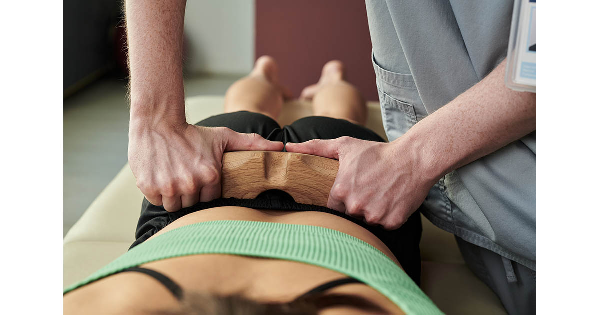
Spinal Decompression for Disc Rehydration
For discs that are symptomatic and still have some viable tissue, motorized spinal decompression is one of the more effective conservative options available. The mechanism is specific: computer-controlled traction creates a carefully modulated distraction force that reduces intradiscal pressure. During that pressure drop, fluid and nutrients are drawn back into the disc space through the endplates.
This does not reverse structural degeneration. A Grade IV disc will not become a Grade II disc through decompression therapy. What decompression can do is reduce the active inflammatory load on a degenerated disc, improve the disc's hydration within its current structural limits, and reduce the nerve irritation that comes from disc height loss. Many patients find that a full course of decompression significantly reduces the daily pain burden even though the structural MRI findings remain unchanged.
Chiropractic Adjustments for Segmental Mobility
Degenerated spinal segments tend to become hypomobile: they lose their normal range of motion as the disc stiffens and the surrounding muscles guard. Restricted segmental motion puts additional stress on adjacent levels, which are then forced to compensate. Over time this compensation pattern spreads the degenerative load.
Chiropractic adjustments restore more normal motion to restricted segments. The effect is not curative for the disc itself, but it interrupts the compensation cascade and can meaningfully reduce the pain that comes from the restricted motion itself. Many of our DDD patients find that regular adjustments reduce the frequency and severity of their flare-ups even if the underlying degeneration remains.
Class IV Laser for Disc and Facet Inflammation
Class IV therapeutic laser penetrates to the depth of the lumbar disc and adjacent facet joints, where it drives mitochondrial activation and accelerates tissue repair at the cellular level. For the inflammatory component of DDD, particularly when there is active facet joint arthritis secondary to the disc degeneration, laser therapy can reduce the inflammatory signal meaningfully. We often combine it with decompression in a sequenced treatment protocol.
Movement and Load Management
Contrary to what many patients expect, the goal is not to stop moving. The disc's nutrition depends on movement. The stabilizing muscles around a degenerated segment need to be strengthened, not rested indefinitely. We work with patients on which movement patterns to modify versus which to maintain, and how to progress activity in a way that supports the disc rather than stressing it further.
Who Gets Better and What to Expect
Most people with symptomatic DDD who pursue consistent conservative care do see meaningful improvement. The research on conservative management of lumbar DDD generally shows positive outcomes for pain and function at 6-12 month follow-up, with the best results in patients who engage actively with their care rather than passively waiting for relief.
The honest caveat: DDD is a structural condition. The underlying disc changes do not reverse. The goal of treatment is to manage the symptomatic expression of those changes, support the health of remaining disc tissue, and slow the progression of secondary changes. Some patients reach a stable baseline with periodic care. Others need ongoing maintenance. A small percentage do eventually pursue surgical options, though this represents a minority even among patients with significant DDD.
What matters most is accurate diagnosis of which specific structures are pain-generating and targeted treatment of those structures, rather than a generic "back pain" protocol applied to everyone with a similar MRI report.
When to Get Evaluated
If you have been told you have degenerative disc disease and you are managing significant daily pain, or if your symptoms are changing in ways you do not understand, getting a proper clinical evaluation is worth doing. MRI findings alone do not drive treatment decisions at our practice. We look at the whole clinical picture: your pain patterns, your functional limitations, your physical exam, and how you respond to targeted provocative testing.
We also see patients who have already tried multiple rounds of conservative care elsewhere and are wondering whether they are missing something. Sometimes a more specific diagnosis of the pain generator reveals a different treatment path that had not been tried.
If you are local to Lakewood Ranch, Bradenton, or Sarasota and you want a straight answer on what your MRI is actually telling you and what to do about it, call us at (727) 213-2982. We can often get you in within 24 hours for a first evaluation.
For more on the conditions that commonly develop alongside DDD, see our back pain conditions page.