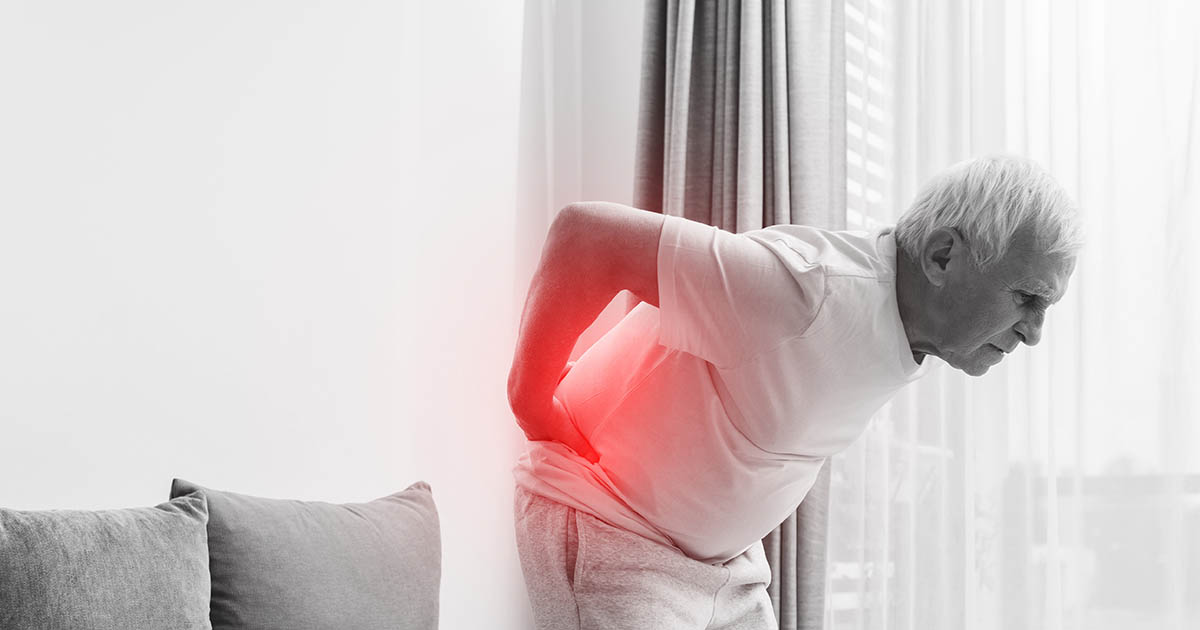
There is a posture pattern so common that physical therapists and chiropractors have a shorthand term for it: APT, short for anterior pelvic tilt. Walk down any office hallway in Lakewood Ranch and you will spot it: belly pushed slightly forward, lower back arched more than normal, buttocks that protrude behind the body's center line. Most people who have it do not know. They just know their lower back hurts.
If your lower back pain is dull, achy, and located at the belt line (not the mid-back, not shooting down the leg), there is a reasonable chance your pelvis is part of the problem. This post explains what anterior pelvic tilt actually is, the muscle pattern that drives it, and how the evaluation process works at our Lakewood Ranch clinic.
What Anterior Pelvic Tilt Actually Means
The pelvis is a ring of bone that sits between your trunk and your legs. Think of it like a bowl of soup. In a neutral position, the bowl is roughly level: the rim at the front (the ASIS, or anterior superior iliac spine) and the rim at the back (the PSIS) are close to the same height.
In anterior pelvic tilt, the front of the bowl tips downward and the back tips upward. Visually, the lower back becomes more arched (hyperlordotic), and the abdomen pushes forward even in people who are not overweight. Some patients describe it as "always sticking my butt out" or "a constant arch I can't get rid of."
The tilt is not a bone problem. The pelvis itself is structurally normal. The problem is in the muscles and soft tissue that hold the pelvis in position. When some of those muscles are persistently short and overactive and others are weak and underused, the pelvis gets pulled into a forward tilt and stays there.
The Muscle Imbalance Behind the Tilt
Four muscle groups drive most anterior pelvic tilt. Two are typically tight; two are typically weak.
Tight and overactive:
- Hip flexors (primarily the iliopsoas and rectus femoris): These muscles run from the front of the lumbar spine and the pelvis down to the thigh. When they stay shortened for hours (sitting at a desk, driving, watching television), they pull the front of the pelvis down and the lower back into extension. Sitting for six to eight hours a day is essentially a long-duration hip flexor stretch in reverse.
- Lumbar erectors: The deep muscles along the lower spine often become chronically tight in people with APT as they work to hold the arched lumbar position upright.
Weak and underused:
- Glutes (gluteus maximus and medius): The glutes are the primary pelvic stabilizers from behind. When they are weak, the pelvis loses its posterior anchor and drifts forward. Many people with APT have strong quadriceps but surprisingly weak glutes, even if they exercise regularly.
- Deep abdominals (particularly the transverse abdominis): The transverse abdominis acts like an internal corset, bracing the pelvis from the front. When it is weak or disconnected from normal movement patterns, the front of the pelvis is unsupported and droops.
This specific four-muscle pattern was described by Czech physiotherapist Vladimir Janda as "Lower Crossed Syndrome." You can read more about the broader pattern in our post on lower crossed syndrome and hip pain, but APT is often how that pattern shows up first.
Why APT Causes Lower Back Pain
Several mechanical processes connect an anteriorly tilted pelvis to back pain.
Facet joint compression. The lumbar vertebrae are stacked with small joints at the back of each level called facet joints. When the lower back is chronically arched (hyperlordotic), the posterior elements of the spine are compressed together. Facet irritation produces a deep, aching pain at the beltline that worsens with standing, walking, or any activity that extends the spine further. Many patients describe it as "hurts when I stand at a party but feels better if I sit and lean forward."
Disc loading changes. The lumbar discs are designed to handle compressive load when the spine is in a neutral or slightly arched position. An exaggerated arch shifts load toward the posterior third of the disc and the facets. Over years, this uneven loading can accelerate disc wear. The most affected levels are typically L4-L5 and L5-S1, which are already the most mobile and most commonly injured levels in the lumbar spine.
Muscle fatigue. The lumbar erectors, already tight, have to work harder to maintain posture when the pelvis pulls them into an arched position all day. This produces a familiar pattern: the back feels fine in the morning, stiffens and aches through the afternoon, and is worst by the end of a workday.
Secondary effects. A tilted pelvis also affects the structures above and below it. The hip flexors, when chronically shortened, can refer pain into the front of the hip or even down the front of the thigh. Some patients with APT develop symptoms that feel like sciatica, but originate from piriformis compression or hip impingement rather than a disc. That distinction matters because the treatment path is different. See our overview of sciatica evaluation and care if your pain radiates below the knee.
How to Do a Rough Self-Check
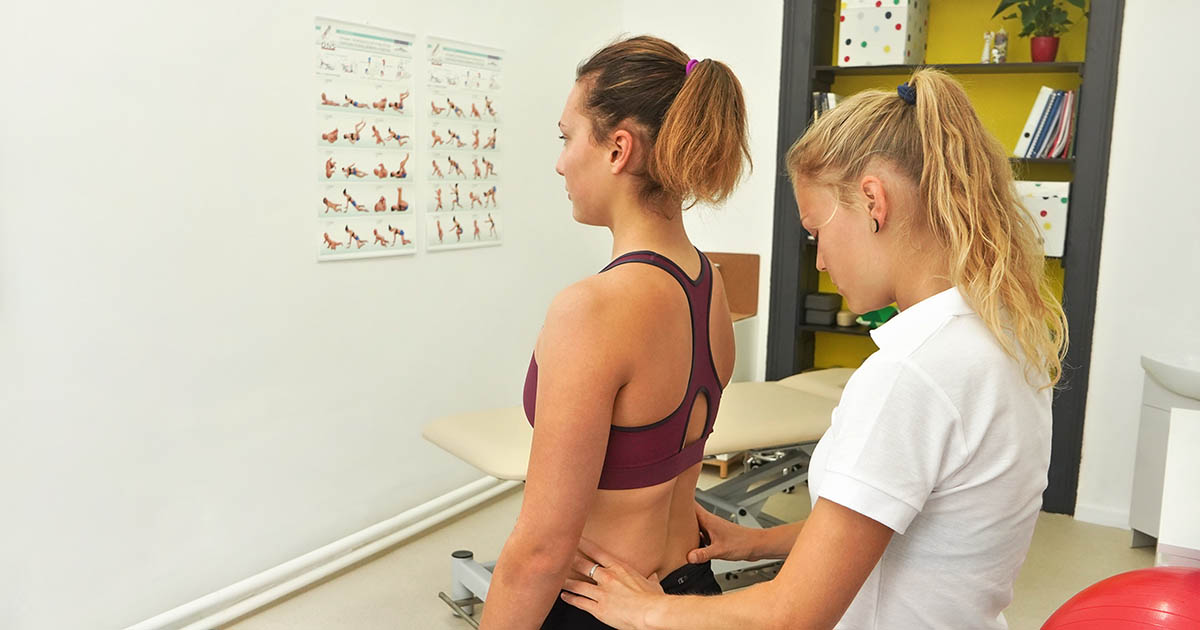
A trained clinician will assess pelvic tilt precisely using anatomical landmarks and posture analysis. But you can do a rough check at home that takes about 30 seconds.
- Stand with your back against a wall, heels about 2 inches from the baseboard.
- Your upper back and the back of your head should touch the wall comfortably.
- Slide your hand into the gap between your lower back and the wall.
- You should be able to slip your hand in, but it should be fairly snug, with maybe one inch of clearance.
- If you can fit a whole fist in the gap, the lumbar arch is likely exaggerated, consistent with anterior pelvic tilt.
This test has real limits. Wall posture does not tell you how you stand when you are tired or moving. And not everyone with APT has obvious hyperlordosis; some have a more subtle tilt. But for a self-screen, a fist-sized gap is worth noting.
"I thought I just had a weak core. Dr. Banman showed me on the posture screen that my pelvis was way forward. The exercises I had been doing were actually making it worse because I kept using my hip flexors instead of my glutes." — many patients describe something very close to this when they finally get a clear picture of what is happening.
Who Tends to Develop Anterior Pelvic Tilt
Certain patterns show up regularly in our Lakewood Ranch clinic:
- Desk workers who sit six or more hours a day. Hip flexors shorten with prolonged sitting. This is the biggest single driver we see. The desk job and lower back pain post covers the broader picture, but APT is a central part of that story.
- People who exercise but skip glute work. Running and cycling keep the hip flexors and quads very active, but neither movement pattern activates the glutes well through a full range. Cyclists in particular often develop significant APT because the hip never reaches full extension on the bike.
- People who had prolonged bed rest or a long recovery. Deconditioning hits the glutes and deep abdominals first.
- New parents who spend a lot of time holding infants on one hip. The compensatory posture loads one side of the pelvis asymmetrically and can establish a tilt over months.
- Patients in the later trimesters of pregnancy. The growing belly shifts the center of gravity forward, which the pelvis accommodates by tilting anteriorly. For most patients this resolves postpartum with appropriate movement, but not always automatically.
What a Clinical Assessment Looks For
When a patient comes in describing persistent lower back aching that does not clearly match a disc pattern, one of the first things we assess is pelvic position and lumbar curve. That evaluation is not just a visual check. It includes:
- Measuring the ASIS-PSIS height difference to quantify the degree of tilt
- Hip flexor length testing (Thomas Test or modified variation)
- Glute strength assessment through single-leg hip extension
- Lumbar range of motion in extension vs. flexion (APT typically hurts with extension and feels better with forward lean)
- Palpation of the lumbar facets and erector spinae for tenderness that correlates with the bony presentation
If the examination supports an APT-driven pain pattern, the treatment focus is different from a disc herniation or a muscle strain. We are working to restore pelvic position through a combination of spinal manipulation (to reduce joint restriction that has built up with the altered mechanics), soft tissue work on the hip flexors and erectors, and clinical guidance on which movements to emphasize versus avoid while the pelvis is retraining.
For patients where the history suggests disc involvement alongside the APT, we will also assess whether non-surgical spinal decompression is appropriate, particularly if there is leg pain or sensory change accompanying the lower back symptoms.
A Note on Exercises (and Why Some Make APT Worse)
Patients with anterior pelvic tilt are often told to "strengthen their core," and they go home and do crunches or planks. Crunches recruit the rectus abdominis but do very little for the transverse abdominis and actually reinforce hip flexor dominance if done improperly. Standard planks, if held with an arched lower back, maintain the very hyperlordotic position driving the problem.
This does not mean exercise is wrong. It means the exercise selection needs to match the specific weakness. Movements that activate the glutes through hip extension (glute bridges, single-leg deadlifts, hip thrusts done with intent) and that train the deep abdominals without hip flexor substitution (dead bugs, pallof press) are the starting point most physios and sports chiros lean toward.
We do not run exercise programs out of our office in the traditional PT sense, but we do give patients very specific guidance on what to do and what to avoid based on the clinical findings. If your back pain is being driven by APT, the last thing we want is for you to spend six weeks doing exercises that make the muscle imbalance worse.
When to Get Evaluated
If your lower back pain fits the description in this post (belt-line ache, worse with standing and extension, better when sitting with a forward lean, present for weeks or months without a clear injury), it is worth getting a structural assessment. Not to confirm that you have anterior pelvic tilt necessarily, but to rule out the patterns that need more urgent attention (disc herniation with nerve compromise, instability, fracture) and to understand what is actually driving the pain before you spend months on treatments aimed at the wrong target.
We see this pattern often enough in our Lakewood Ranch clinic that we have a standard evaluation pathway for it. Most patients leave the first visit with a clear answer about their pelvic alignment and a plan that makes sense for their specific findings.