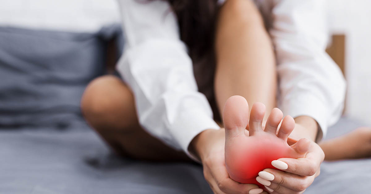
Pain at the back of the heel is one of the more overlooked complaints we see in our Lakewood Ranch clinic. Patients often wait weeks or months before seeking care because the pain eases once they warm up and walk around. That misleads people into thinking the problem is going away on its own. It usually is not. The tendon is accumulating load damage faster than it is repairing itself, and the window for conservative care closes if that pattern runs long enough.
Achilles tendinitis is not just a runner's injury. In our patient population here in the Lakewood Ranch area, it shows up most often in pickleball players, walkers who increased their mileage, golfers on a new routine, and people who spent a Florida summer mostly indoors and then returned to activity when the heat broke. Any pattern that spikes tendon load faster than the tissue adapts is enough.
What the Achilles tendon actually is (and why it struggles to heal)
The Achilles tendon is a thick rope of densely packed collagen fibers connecting the two calf muscles (gastrocnemius and soleus) to the heel bone (calcaneus). In running, it transmits forces six to eight times body weight. In walking, it carries two to three times body weight at push-off. It is the strongest tendon in the human body by a wide margin, which is part of why it takes real cumulative overload to injure it.
Here is the structural catch: the mid-portion of the Achilles is largely avascular. It receives poor blood supply compared to the muscle belly above it or the bony attachment below. Collagen turnover is slow. This means when micro-tears accumulate (the typical mechanism of overuse tendinopathy), the tissue cannot repair at the same rate it breaks down. The result is a degenerating tendon that becomes progressively thickened, irregular, and pain-sensitive before it ever reaches a frank tear.
The clinical term has shifted over the years. "Tendinitis" implies active inflammation, which is present in the early reactive phase. "Tendinosis" or "tendinopathy" more accurately describes the degenerative, disorganized collagen pattern seen in chronic cases. In practice, many clinicians use tendinitis because it is what patients recognize, but the underlying biology matters for choosing treatment.
Insertional versus mid-portion: two different problems
The location of your pain matters a great deal for treatment. Achilles tendinopathy divides into two clinically distinct presentations.
Mid-portion Achilles tendinopathy involves pain 2 to 6 centimeters above the heel bone, roughly in the middle of the cord. This is the most common form in recreational athletes in their 30s to 60s. It correlates with high repetitive loading, tight calves, and rapid increases in activity. Tenderness with direct palpation of the tendon mid-substance is the hallmark finding. The area is often visibly or palpably thickened.
Insertional Achilles tendinopathy involves pain directly at the heel bone insertion, sometimes accompanied by a Haglund deformity (a bony prominence at the posterior heel, sometimes called "pump bump"). It tends to occur in an older patient group, is associated with stiffness rather than swelling, and is often aggravated by the heel tab of a shoe pressing into the tendon. Treatment protocols for insertional tendinopathy differ from mid-portion protocols, particularly in the use of heel drops and the angle at which loading exercises are performed.
Getting this distinction right matters because a protocol that works well for one type can aggravate the other.
Why start-up pain and morning stiffness are diagnostic clues
The characteristic pain pattern of Achilles tendinopathy is morning start-up pain: intense discomfort with the first several steps out of bed, easing over 5 to 15 minutes as the tendon warms and fluid distributes. The same pattern repeats after sitting for a prolonged period and then rising to walk.
This can be confused with plantar fasciitis, which has nearly identical morning-step pain. The key distinguishing feature is location: plantar fasciitis pain is at the bottom of the heel and arch (the plantar fascia insertion). Achilles pain is at the back of the heel or in the cord above it. If you are pressing on the back of your heel or the calf cord itself and that is where it hurts, the plantar fascia is not the primary driver. For a detailed breakdown of the plantar fascia pattern, see our post on foot pain that gets worse in the morning.
Start-up pain that eases with movement but returns after activity is not a good sign that things are healing. It is a sign that the tendon is irritated but not yet at a threshold to produce resting pain. The window for conservative care is still open, but it requires action.
The five most common drivers of Achilles tendinopathy in active adults
Achilles tendinopathy almost always has a training or load error at its root, combined with one or more structural factors that make the tendon more vulnerable. The most common drivers we see:
- Training load spike: Returning to activity after a break, adding a second sport, doubling pickleball sessions, or increasing walk distance in a short window. The tendon adapts more slowly than cardiovascular fitness. Your lungs clear the 5-mile mark long before your Achilles tolerates it.
- Calf weakness or tightness: The gastrocnemius and soleus absorb push-off forces before they reach the tendon. If those muscles are weak or chronically tight, more raw force transfers directly to the cord. Tight calves also place the tendon under higher resting tension, which compounds cyclic loading stress.
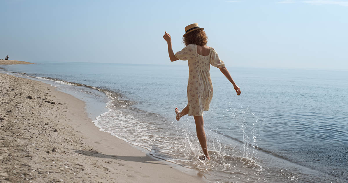
- Overpronation: When the foot rolls inward at push-off, the Achilles does not pull in a straight line. The medial cord takes disproportionate load and a twisting moment the tendon is not designed for. This pattern is especially common in people who walk extensively on beach sand or uneven surfaces, which is another reason this injury appears frequently in coastal Florida.
- Footwear change: Switching from a cushioned running shoe to a minimalist flat shoe or going barefoot inside reduces the heel-toe drop and loads the Achilles through a greater range than it has adapted to. The flip side also applies: a worn-out shoe with a collapsed heel counter no longer provides adequate support.
- History of corticosteroid injection near the tendon: Cortisone injected directly into or immediately adjacent to the Achilles weakens collagen temporarily. If significant activity followed within weeks, micro-structural damage can accumulate rapidly. This is a documented risk factor for partial and complete tears and is one reason steroid injection into the Achilles is generally avoided by informed clinicians.
When tendinopathy becomes a partial or complete tear: red flags
Most Achilles tendinopathy cases remain at the tendinopathy stage and respond to conservative care if addressed appropriately. A subset, particularly those with longstanding degeneration or an acute high-load event on top of a chronically irritated tendon, progress to partial or complete rupture.
Signs that a tear may have occurred:
- An audible or palpable pop at the back of the heel during activity
- Sudden inability to push off the foot or stand on the toes of the affected leg
- Significant swelling and bruising around the lower calf and heel
- A positive Thompson test (squeezing the calf does not produce foot plantarflexion)
- A palpable gap in the tendon cord
Any of these findings warrants immediate orthopedic evaluation and imaging, not conservative chiropractic or manual therapy. We do not treat acute Achilles ruptures conservatively, and if we see these signs on exam we refer patients out immediately.
What conservative care actually accomplishes
A well-structured conservative program for Achilles tendinopathy works toward three goals: reduce tendon irritation, rebuild load tolerance, and correct the upstream biomechanical driver.
Reducing irritation means temporarily modifying the activities that provoked the flare while keeping the tendon in controlled, graded load. Complete rest is counterproductive: tendons need mechanical stimulus to signal collagen remodeling. The goal is finding the load level where symptoms are manageable but tissue adaptation is still occurring.
Rebuilding load tolerance centers on eccentric and heavy slow resistance exercises for the calf-Achilles unit. Eccentric heel drops (lowering slowly on one foot from a step) are among the most well-studied conservative interventions for mid-portion Achilles tendinopathy. They place the tendon under controlled elongation load, which appears to stimulate better collagen organization than concentric-only loading.
Correcting upstream drivers is where chiropractic and structural work is most relevant. If overpronation, ankle joint restriction, hip weakness, or gait asymmetry is loading the Achilles unevenly, the tendon will not durably recover until those mechanics are addressed. Chiropractic adjustments to the ankle, subtalar joint, and lower extremity kinetic chain can restore the joint mechanics that the tendon depends on. Functional orthotics may also be part of the picture when structural foot issues are a significant contributor.
For beach walkers and outdoor exercisers in Lakewood Ranch, we also work through activity modification specific to soft-sand walking, which places the Achilles under a substantially different loading pattern than pavement. See our post on why beach walking hurts the legs more than regular walking for a deeper breakdown of that mechanism.
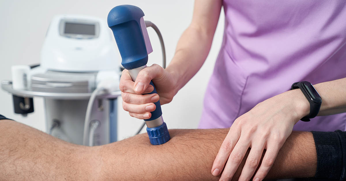
Shockwave and Softwave therapy for tendon tissue repair
For cases that are not responding to loading-based conservative care alone, acoustic wave therapy has emerged as one of the better-studied adjunct interventions for chronic tendinopathy.
Shockwave therapy delivers high-energy acoustic pulses to the tendon. The proposed mechanisms include stimulating neovascularization (new blood vessel formation in the avascular mid-portion), disrupting calcific deposits at the insertion, and triggering a controlled cellular repair response. For insertional Achilles tendinopathy with calcification, shockwave has a reasonably strong evidence base. For mid-portion tendinopathy, results are more variable but consistently better than placebo in better-designed trials.
Softwave therapy uses a different wave delivery system, producing broader, lower-peak-pressure waves that penetrate tissue with less discomfort than traditional focused shockwave. In our experience at the Lakewood Ranch clinic, patients tolerate Softwave sessions well, and it is particularly useful for patients who have found standard shockwave too uncomfortable or who have concurrent soft-tissue involvement around the tendon.
Neither shockwave nor Softwave is a substitute for the loading program. They work best as part of a combined approach where the tissue is simultaneously being mechanically reconditioned through graded exercise.
A typical course for Achilles tendinopathy at our clinic involves an initial evaluation to confirm the diagnosis and identify the specific drivers, followed by a structured program combining manual therapy, loading progression, and in appropriate cases, acoustic wave therapy. Many patients see meaningful improvement within 6 to 12 weeks with consistent adherence to the program.
When to seek evaluation
Achilles tendinopathy responds better to care when it is addressed early, before the collagen architecture degrades substantially. The following situations warrant an evaluation rather than continued self-management:
- Pain that has been present for more than 4 to 6 weeks without clear improvement
- Pain that is interfering with daily walking, not just exercise
- A visible or palpable thickening of the tendon cord
- Pain that worsens over the course of an activity rather than warming up and improving
- Any history of corticosteroid injection near the tendon in the past 3 to 6 months
If you are in the Lakewood Ranch, Bradenton, or Sarasota area and recognizing this pattern, call us at (727) 213-2982 or book online below. We evaluate and treat Achilles tendinopathy as part of our broader musculoskeletal program, with access to the full range of acoustic wave and manual therapy tools in-house.