You wake up, swing your legs over the side of the bed, and the moment your foot hits the floor you immediately know the day is starting badly. Sharp pain shoots through your heel or arch. You limp to the bathroom. By the time you have been on your feet for ten minutes, it has backed off to a dull ache. By mid-afternoon, you almost forget it was there. Then you sit down for a long meeting, stand up, and it spikes again.
If that sequence sounds familiar, you are not imagining a pattern. That exact pattern, severe pain with the first steps and some relief after warm-up, has a specific mechanical explanation. Understanding it is the first step toward actually addressing it rather than just waiting it out.
Why morning is the worst time for foot pain
When you sleep, your foot spends several hours in a plantar-flexed position: toes pointing slightly downward, the muscles and soft tissues on the underside of the foot shortened and relaxed. The plantar fascia, a thick band of fibrous tissue running from the heel bone to the base of the toes, shortens along with everything else.
When you stand and take your first step, you abruptly load that shortened tissue with your full body weight. If there is existing inflammation, micro-tearing, or structural irritation in the plantar fascia or the structures around it, that first step forces a rapid stretch from a shortened overnight position. The result is a sharp, stabbing sensation at the heel or arch.
As you continue walking, circulation increases, the tissue warms, and the initial stiffness gradually eases. That pattern, severe with first steps, improving over minutes of movement, is one of the most characteristic features of plantar fasciitis. But it is not universal across all conditions that produce foot pain, which is why the specific pattern matters when working toward a diagnosis.
The most common cause: plantar fasciitis
Plantar fasciitis is the most frequently diagnosed cause of morning heel and arch pain. It involves the plantar fascia, the thick fibrous band that supports the arch of the foot and absorbs a significant portion of the mechanical load every time your heel strikes the ground.
Despite the name, plantar fasciitis is less a true inflammatory condition and more a degenerative one. The tissue develops micro-tears at its attachment point near the heel bone (the calcaneus) when it is repeatedly loaded beyond its ability to recover. Those tears accumulate, the tissue becomes structurally disrupted, and pain results when it is stressed.
In Southwest Florida, this condition is extremely common. Hard tile and concrete floors in homes and businesses, warm weather that leads to more time in flat sandals and flip-flops, and the general activity level of Lakewood Ranch residents (pickleball, walking trails, gardening, golf) all contribute to the fascial load that drives plantar fasciitis.
Contributing factors that increase risk:
- Increased activity volume without adequate conditioning buildup
- Long periods of standing on hard floors (teachers, kitchen workers, retail staff)
- Unsupportive footwear: flip-flops, flat canvas shoes, worn-out athletic shoes
- Tight calf muscles or Achilles tendon, which limits ankle dorsiflexion and increases fascial load
- Flat feet or high arches, both of which alter how the fascia distributes impact
- Excess body weight, which increases the compressive and tensile forces on the fascia with every step
- Sudden changes in walking surface (from carpeted surfaces to beach sand or pavement)
For a full clinical picture of how plantar fasciitis is evaluated and managed, see our plantar fasciitis page.
Other structures that produce morning foot pain
Plantar fasciitis is the most common diagnosis, but not the only one. Several other conditions produce a similar pattern, and they require different approaches:
Heel fat pad syndrome
The heel pad is a specialized cushioning structure that protects the calcaneus during impact. With age or sustained high-impact activity, this pad can thin or partially fragment. Pain is more diffuse than plantar fasciitis and tends to worsen throughout the day rather than ease after warm-up. There is no effective way to rebuild lost fat pad tissue, so management focuses on off-loading and external cushioning.
Achilles tendinopathy
The Achilles tendon attaches to the back of the heel bone. Tendinopathy (degeneration within the tendon itself) produces pain that is worst with first steps in the morning and with stair climbing. The location is the back of the heel, not the bottom, which is often enough to distinguish it from plantar fasciitis on palpation. Achilles tendinopathy responds to different loading protocols than plantar fasciitis does.
Tarsal tunnel syndrome
Like carpal tunnel syndrome in the wrist, tarsal tunnel syndrome involves compression of the posterior tibial nerve as it passes through a fibrous canal on the inner side of the ankle. It produces burning, tingling, or stabbing pain in the heel and arch. The neurological quality of the pain, and the fact that it may radiate into the toes, distinguishes it from purely mechanical heel pain. If your morning foot pain includes tingling, burning, or numbness, nerve involvement needs to be evaluated.
Stress fractures
A calcaneal stress fracture produces heel pain that is severe with weight-bearing and does not ease with walking. It typically comes after a significant increase in activity (such as starting a running program or doing extensive hiking) and localizes to the body of the heel rather than the plantar surface. If the pain is acute, severe, and does not respond to the standard morning warm-up, imaging is appropriate.
Inflammatory arthritis
Conditions like rheumatoid arthritis, psoriatic arthritis, and ankylosing spondylitis can produce foot and heel pain that is most pronounced in the morning and improves with movement. The distinguishing feature is duration: inflammatory morning stiffness typically lasts over an hour and is often bilateral (both feet). Mechanical plantar fasciitis usually peaks in the first five to ten minutes and is more commonly unilateral.
The morning warm-up test is useful but not definitive. If your morning pain eases within ten minutes of walking, that pattern fits a mechanical tissue injury well. If it persists past 30-60 minutes, or if both feet are involved symmetrically, that raises the possibility of a systemic inflammatory condition that warrants a different kind of evaluation.
Why rest makes it worse, not better
This is one of the most frustrating realities for patients with plantar fasciitis. "I slept eight hours. Why is it worse than it was at the end of yesterday?"
Here is the mechanism. During the day, the micro-tears in the plantar fascia are being repeatedly stressed, but they are also receiving continuous blood flow and mechanical stimulation that drives the repair process. The tissue is actively trying to heal even as it is being loaded.
During sleep, circulation to the foot decreases, the tissue cools and shortens, and the repair activity slows. The small amount of healing that was progressing essentially pauses. By morning, the tissue has "set" in that shortened, relatively stiff position. When you then load it fully with your first step, you are stressing tissue that is at its stiffest and most vulnerable point of the day.
This is the physiological basis for night splints, which hold the foot in mild dorsiflexion overnight. By keeping the plantar fascia at a slightly lengthened position during sleep, the morning load does not require as dramatic a stretch transition. Many patients notice a meaningful reduction in first-step pain within a few nights of consistent splint use.
Red flags that change the evaluation
Most morning foot pain is mechanical, benign in origin, and responds well to conservative care. A few patterns point to something that needs faster attention:
- Pain that does not ease at all with walking and worsens as the day goes on
- Significant swelling, bruising, or heat in the foot or ankle
- Fever alongside foot pain (possible infection or systemic condition)
- Burning, tingling, or numbness that radiates into the toes
- Bilateral heel pain that started at the same time in both feet
- Pain that wakes you from sleep (mechanical pain typically settles at rest)
- Acute onset after a fall, heavy impact, or change in footwear with no gradual build-up
Any of these patterns warrants evaluation before initiating a soft-tissue treatment protocol. If the clinical picture suggests a stress fracture, nerve compression, or systemic inflammatory process, the appropriate next step is imaging or referral, not manual therapy or laser treatment.
What a clinical evaluation looks at
Patients often arrive having already tried the obvious: rest, ice, stretching their calf on the stairs. The pain came back as soon as they resumed normal activity. A structured evaluation goes further than those self-management trials.
For morning foot pain, a thorough exam typically includes:
- Pain location: medial calcaneal tubercle (bottom of heel), Achilles insertion (back of heel), midarch, or diffuse
- Pattern history: duration of symptoms, what makes it better or worse, bilateral or unilateral
- Provocation testing: palpation, the Windlass test, dorsiflexion stretch
- Ankle and subtalar joint range of motion
- Gastrocnemius and soleus flexibility assessment
- Gait observation: how the foot loads through heel strike, midstance, and toe-off
- Foot posture index: degree of pronation or supination at rest and during movement
- Neurological screen: light touch, vibration sense, Tinel sign at the tarsal tunnel if nerve involvement is suspected
The goal is not to simply name the condition. It is to identify which specific structure is the primary driver so care targets the right thing from the start. "Heel pain" is not a treatment target. "Degenerative plantar fasciosis at the medial calcaneal tubercle with limited Achilles flexibility as a primary load driver" is.
Treatment approaches for plantar fasciitis
For the most common presentation, mechanical plantar fasciitis, care combines reducing the immediate load on the irritated tissue while improving the tissue's long-term capacity to handle that load.
Mechanical load modification
This means evaluating footwear first. A flat flip-flop provides almost no arch support and allows the plantar fascia to stretch excessively with every step. Walking barefoot on tile floors at home, especially in the morning, is one of the most consistent aggravating factors we hear about. A supportive athletic shoe or a firm orthotic inside a structured shoe can meaningfully reduce the per-step fascial load during the recovery period.
Calf and Achilles flexibility
Tight gastrocnemius and soleus muscles limit how much the ankle can dorsiflex (flex upward) during walking. When ankle dorsiflexion is restricted, the plantar fascia is recruited to make up the difference in flexibility during push-off. More recruitment means more cumulative load and more micro-tearing. Specific stretching protocols targeting these muscles, particularly a bent-knee calf stretch that isolates the soleus, are among the most consistently supported interventions in the clinical literature on plantar fasciitis.
Shockwave therapy
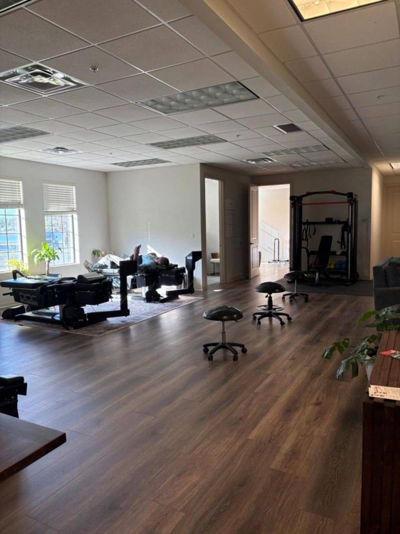
For plantar fasciitis that has persisted despite activity modification and stretching (typically three or more months), shockwave therapy applies focused acoustic energy to the damaged tissue at the fascial insertion. Rather than simply "breaking down scar tissue," shockwave appears to stimulate a localized healing response in tissue that has stalled in a chronic degenerative cycle, essentially re-initiating the repair process. Our Lakewood Ranch clinic uses shockwave as part of a structured care plan for appropriate candidates. See our shockwave therapy page for a fuller explanation of how the technology works and who tends to benefit most.
Class IV laser therapy
Laser therapy at Class IV wavelengths drives photobiomodulation at the cellular level: it increases ATP production in damaged tissue and modulates inflammatory cytokine activity. For plantar fascia injuries, this can accelerate tissue-level healing and reduce pain in the early and mid phases of care. It is particularly useful in combination with mechanical interventions, since reducing the inflammatory component makes the tissue more responsive to loading-based rehabilitation. Our Class IV laser page explains how this differs from cold or low-level laser, which operates at substantially lower power output and different tissue penetration depths.
Joint mobilization
The ankle, subtalar joint, and midfoot joints all contribute to how the foot loads during walking. Restriction in any of these joints alters the mechanical environment of the plantar fascia and can maintain or worsen the load pattern driving the injury. Manual mobilization to restore motion at restricted joints is often part of a complete care plan, particularly when gait analysis shows compensatory patterns.
Night splints
As described earlier, keeping the foot in mild dorsiflexion overnight prevents the tissue from shortening and setting during sleep. Night splints are not comfortable for everyone, and compliance is variable. For patients who can tolerate them, they are often the intervention that produces the fastest reduction in morning pain severity.
What to do right now before your evaluation
If you are dealing with morning foot pain and have not yet been evaluated, a few steps are worth implementing immediately:
- Stop walking barefoot on hard floors, especially in the morning. Keep supportive footwear next to the bed and put it on before your first step.
- Stretch before you stand. While still lying down, flex your foot up and down 15-20 times and pull your toes back toward your shin with your hand. Pre-loading the tissue before bearing weight reduces the first-step pain spike.
- Apply ice after activity, not before. Ice after walking reduces local inflammation. Using it as a pre-activity warm-up does not address the mechanical issue and can delay healing if over-applied.
- Evaluate your footwear. Athletic shoes need replacing roughly every 400-500 miles of use. Worn-out midsoles provide minimal support regardless of how the shoe looks from the outside.
- Avoid complete rest. Total cessation of walking allows the tissue to shorten and stiffen further. Light walking on a supportive surface maintains circulation and mechanical stimulus for healing better than full rest.
These are management strategies, not fixes. They can reduce daily pain while you work toward evaluation and care. They do not address the underlying structural or mechanical driver, and for most people with true plantar fasciitis, self-management alone does not resolve the problem.
When to get evaluated
Morning foot pain that has lasted more than two to four weeks without improving, that limits your ability to walk normally, or that keeps returning after brief improvement is worth a structured clinical evaluation. The longer degenerative plantar fasciitis is allowed to progress without addressing the mechanical driver, the more tissue disruption accumulates and the more involved the recovery tends to be.
Early evaluation also helps rule out the conditions listed above, particularly if the pain pattern does not fit the classic plantar fasciitis profile. Treating a tarsal tunnel problem like a fascial problem, or continuing with conservative care for a stress fracture, causes delay and additional damage.