Most people have heard of a herniated disc. Fewer have heard of facet joint syndrome, even though facet joints are involved in a significant portion of chronic spinal pain cases. Studies in the pain medicine literature estimate that lumbar facet joints account for roughly 15 to 45 percent of chronic low back pain, depending on the population studied. Cervical facets are implicated in up to 60 percent of chronic neck pain after whiplash. These are not small numbers.
So why does this diagnosis get missed so often? Partly because the pain pattern overlaps with other conditions. Partly because routine imaging does not always make it obvious. And partly because "facet joint syndrome" is a mouthful that patients and some providers skip past on their way to more familiar diagnoses.
This post covers what facet joints are, how their breakdown produces pain, how to recognize the pattern, and what conservative care at a chiropractic and wellness clinic can do to address it.
What Are Facet Joints and Why Do They Matter
The spine is not a single solid structure. It is a stacked column of 24 moveable vertebrae connected at three points per level: the disc in front, and two facet joints (also called zygapophysial joints, or z-joints) positioned on the left and right at the back. Every time you rotate, extend, or laterally bend your spine, the facet joints guide and limit that movement. They keep the vertebrae from translating out of position and protect the spinal cord and nerve roots from excessive mechanical stress.
Like any synovial joint in the body, such as a knee or a finger knuckle, facet joints have cartilage surfaces, a synovial lining that produces lubricating fluid, and a fibrous joint capsule. The capsule is richly innervated by small nerves called medial branches, which is what makes the joints capable of generating significant pain when they become inflamed or degenerate.
Facet joint syndrome is the clinical term for pain that originates from one or more of these joints due to inflammation, capsular irritation, or cartilage degeneration. It is a musculoskeletal diagnosis, not a structural emergency in most cases, but it can become a persistent, quality-of-life-limiting problem when it goes unrecognized and the underlying mechanics are not addressed.
Why Facet Joint Pain Gets Missed So Often
There are several converging reasons why this diagnosis falls through the cracks.
The pain pattern is not as dramatic as classic sciatica. A herniated disc compressing a nerve root produces sharp, electric pain that radiates clearly down the leg, often past the knee, with a predictable dermatomal distribution. Facet joint pain is usually duller and more local: it stays near the spine, refers into the buttock or hip in a vague, achy pattern, and rarely travels below the knee. Patients often describe it as a "muscle ache" or "tightness" rather than a sharp nerve pain, which leads to days of stretching and heating pads before they see a provider.
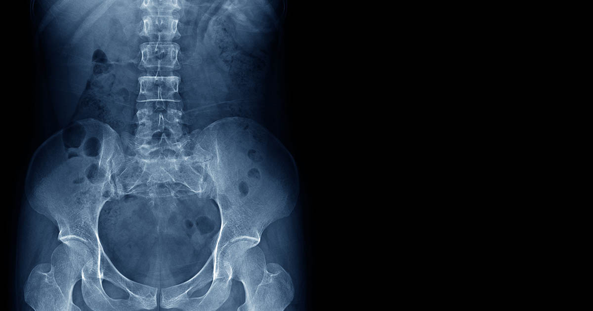
Standard imaging underreports it. X-rays show bone and can reveal facet joint space narrowing or osteophyte formation in more advanced cases, but earlier-stage facet irritation will not be visible. MRI shows more soft tissue detail, but the radiologist's report may note only "mild degenerative changes throughout" without calling out the facet joints specifically as the dominant pain generator. A patient can have clinically significant facet syndrome and receive an imaging report that sounds relatively benign.
Disc and facet pathology coexist frequently. The disc and the facet joints at each spinal level form a three-joint complex that loads and degenerates together. When a disc loses height, the facet joints above and below it change their loading angle and begin to carry more mechanical stress. The result is often a mix of disc and facet changes on imaging, and clinical attention tends to focus on the disc, leaving the facet contribution unaddressed.
The Classic Signs That Point to the Facet Joints
Pain patterns matter. Facet joint syndrome tends to produce a recognizable cluster of clinical findings that differ meaningfully from disc-driven pain:
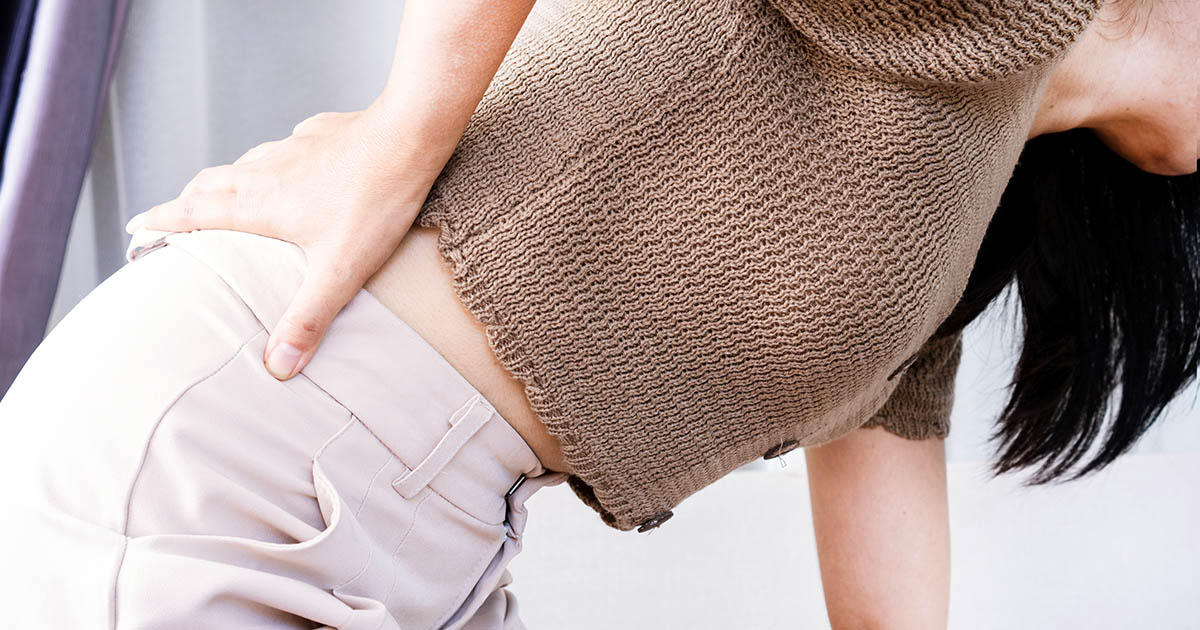
- Pain that is worse with extension and rotation. Facet joints are most compressed when the spine is in extension, meaning arching backward, standing tall for long periods, walking on flat surfaces for extended time, or sleeping face-down. Twisting aggravates them further. Bending forward opens the facet joint space and typically provides relief. Sitting is often more comfortable than standing.
- Tenderness directly over the facet joints. The lumbar facet joints sit approximately 1.5 to 2 centimeters lateral to the spinous processes (the bony bumps you feel down the center of your back). Firm pressure over those spots is reliably painful when the joints are inflamed. Cervical facets are accessible at similar lateral-to-spinous positions in the neck.
- Referred pain to the buttock, hip, or upper thigh without true radiculopathy. Lumbar facet joints refer pain in a predictable but non-dermatomal pattern: the lower lumbar joints tend to refer into the buttock, posterior hip, and proximal thigh. This can feel like hip bursitis or a pulled glute, and it occasionally gets confused with early sciatica. The key distinction: it rarely radiates past the knee, and it does not have the sharp, electric, dermatomal quality of true nerve root compression.
- Cervical facet pain that feels like a "tension headache" or shoulder ache. Upper cervical facet involvement (C2-C3, C3-C4) commonly refers to the base of the skull, the occiput, and behind the ear. Mid-cervical facets refer into the neck and shoulder in a pattern that is frequently attributed to muscle tension. Many patients in our office who have been told they "just carry stress in their neck" for years have cervical facet involvement that has never been directly addressed.
- Morning stiffness that improves with gentle movement, then worsens again with prolonged loading. Unlike disc pain, which often peaks with extended rest and improves once the disc rehydrates with movement, facet pain can have a more variable pattern: it may ease with a morning walk but return after a long afternoon on your feet or a multi-hour car ride.
If your back pain is significantly better when you sit and lean forward, and worse when you stand or walk, the facet joints are high on the differential. That extension-intolerant pattern is one of the most reliable clinical clues we have.
What Causes Facet Joints to Degenerate
Facet joints are designed to tolerate a lifetime of mechanical loading, and for many people they do. But several factors accelerate the breakdown:
Cumulative postural load over years. Extended sitting with a flattened lumbar curve shifts load away from the discs and onto the posterior structures, including the facet joints. For desk workers and drivers, this is a daily and sustained mechanical stress. The Lakewood Ranch and Sarasota area has a significant population of commuters on I-75 and office workers who sit for six to eight hours a day. We see the cumulative effects consistently.
Disc degeneration as a trigger. When a disc loses height, it changes the biomechanics of the entire three-joint complex at that level. The facet joints are forced into a slightly different angle and begin bearing a disproportionate share of compressive load. Facet arthritis and disc degeneration often advance in parallel, which is why the two are so frequently found together on imaging.
Prior injury to the joint capsule. A hyperextension event, a sports injury, a fall, or even a rear-end collision can overstretch or micro-tear the facet joint capsule. The capsule has a rich nerve supply, and once sensitized, it can remain irritable long after the acute injury appears to have resolved. This is part of why some patients struggle with lingering back or neck pain for months after an accident even when their disc findings are unremarkable. For patients with auto injuries, documenting facet joint involvement is part of comprehensive injury care. Our clinic coordinates with attorneys when documentation is needed for a PIP or personal injury claim.
Repetitive extension-loaded activities. Overhead work, certain swimming strokes, back extensions in the gym, and occupations requiring frequent lumbar extension all add repetitive compressive stress to the facet joints. Over time, the cartilage surfaces thin, the capsule stiffens, and the joint develops a chronic low-grade inflammatory state.
How Facet Joint Syndrome Is Evaluated
There is no single test that definitively diagnoses facet joint syndrome in a clinical office setting. What matters most is the combination of history, physical findings, and, when appropriate, imaging and diagnostic procedures.
In our Lakewood Ranch clinic, evaluation starts with a detailed history: where does it hurt, what makes it better or worse, what is the quality and referral pattern of the pain, and how does the pattern compare to the signs listed above. Physical examination includes palpation of the facet joint regions, assessment of spinal range of motion (specifically extension and rotation), and provocative testing to distinguish facet-driven referred pain from true radiculopathy.
Imaging is ordered when the clinical picture warrants it. X-rays can reveal facet joint space narrowing and hypertrophic changes in advanced cases. MRI provides additional detail on cartilage, joint fluid, and the relationship between facet pathology and adjacent disc and neural structures.
The most specific available test is a diagnostic medial branch block, in which a small volume of local anesthetic is injected under fluoroscopic or ultrasound guidance to temporarily numb the nerves supplying the facet joints. If pain is substantially relieved for the expected duration of the block, that confirms the facet joints as the primary pain generator. This is typically arranged through a pain management physician or interventional radiologist. Our clinic coordinates referrals when this level of diagnostic specificity is needed, and we continue conservative care before and after the procedure.
What Conservative Care Can Actually Do
Facet joint syndrome responds well to conservative care when the approach is matched to the mechanism. Here is what we see work in practice at our clinic and what the clinical literature supports:
Chiropractic adjustments targeted to hypomobile segments. When a facet joint becomes chronically irritated, the surrounding capsule and adjacent musculature often tighten reactively, restricting movement and perpetuating the pain cycle. Specific mobilization or manipulation techniques restore motion to the restricted segment. This is not about "cracking" the spine indiscriminately; it is about identifying the exact levels where movement is restricted and applying appropriately calibrated force to restore that motion. For many patients, the relief after a well-targeted adjustment is immediate, though multiple sessions are usually needed for lasting improvement. Our chiropractic adjustment care is always preceded by a thorough assessment of which segments need to be addressed and how.
Class IV laser for joint capsule inflammation. Photobiomodulation using a Class IV therapeutic laser delivers light energy at wavelengths that penetrate to the depth of the facet joint capsule, where it promotes cellular repair, reduces inflammatory mediators, and has a documented analgesic effect on sensitized joint tissue. Cold laser units lack the power density to reach the relevant tissue depths in a clinical timeframe. Our Class IV laser protocol typically runs 8 to 12 sessions for facet-driven inflammation, and many patients report meaningful reduction in background aching within the first few visits.
Spinal decompression when disc involvement is present. Because facet degeneration and disc degeneration so often coexist, many facet syndrome patients have a disc component that benefits from spinal decompression therapy. Decompression creates a negative intradiscal pressure gradient that promotes disc rehydration and can reduce the secondary mechanical overload transferred to the facet joints. When imaging shows both facet and disc changes, combining decompression with manual therapy and laser often produces better functional outcomes than any single approach alone.
Postural and movement correction. The goal of rehabilitation is to modify the mechanical loads that are perpetuating facet irritation. For lumbar facet syndrome, this typically means improving hip mobility (tight hip flexors drive the pelvis into an anterior tilt that increases lumbar extension), strengthening the deep stabilizers of the spine without repeatedly loading it in extension, and correcting workplace ergonomics. For cervical facet syndrome, forward head posture correction is central: every inch of forward head carriage increases the effective compressive load on the posterior cervical structures by roughly 10 pounds.
Medial branch radiofrequency ablation (RFA) for refractory cases. When conservative care produces partial improvement but significant residual pain persists despite an adequate course of treatment, referral to a pain management physician for diagnostic blocks and possible RFA is appropriate. RFA uses heat generated by radiofrequency energy to disrupt the medial branch nerves supplying the facet joints, providing pain relief that typically lasts 12 to 18 months in good candidates before the nerves regenerate. We refer patients for this when the clinical picture warrants it and co-manage their rehabilitation around the procedure.
What Does Not Help (and Can Make Things Worse)
Extended rest makes facet joint syndrome worse. Bed rest allows surrounding musculature to decondition, reduces synovial fluid circulation within the joint, and does nothing to address the mechanical loading patterns that are perpetuating the problem. Short rest periods during acute flares are reasonable. Prolonged rest is not a treatment strategy.
Extension-biased exercise protocols developed for disc problems, such as the McKenzie extension press-up series, are contraindicated for primary facet syndrome. Extension is the movement that compresses and loads the facet joints. Applying it as a treatment will reliably aggravate the condition.
Long-term reliance on NSAIDs can mask pain without addressing the underlying mechanics, and extended NSAID use carries known gastrointestinal and cardiovascular risks. A short course of anti-inflammatories under physician guidance is reasonable for acute flares. Using them as the sole long-term strategy is not.
When to Get Evaluated Right Away
Most facet joint syndrome is a chronic but manageable musculoskeletal condition. The following findings are reasons to seek evaluation promptly rather than continuing self-management:
- New onset of leg weakness, foot drop, or significant changes in bladder or bowel function. These suggest nerve root or spinal cord involvement that needs urgent assessment.
- Rapidly worsening neurological symptoms rather than the typical chronic slow progression of facet arthritis.
- Back or neck pain accompanied by fever, night sweats, or unexplained weight loss. These raise the possibility of infection or malignancy and need medical evaluation before attributing the pain to facet joints.
- Pain that wakes you consistently from sleep and does not respond to any position change. Most mechanical spinal pain, including facet syndrome, finds at least one position of partial relief. Pain that is truly positional-independent warrants further investigation.
For a comprehensive overview of the different sources of spinal pain and how they are evaluated, see our back pain conditions page.
Getting an Answer at Our Lakewood Ranch Clinic
If your back or neck pain fits the pattern described above, the first step is a clear clinical assessment that identifies where the pain is actually coming from. Many patients we see have been stretching the wrong structure, icing the wrong area, and taking medication for a diagnosis that was never precisely established. Getting the diagnosis right is what makes the treatment right.
At Spine and Wellness Center Lakewood Ranch, Dr. Banman evaluates the full picture: disc, facet joints, sacroiliac joints, and the adjacent soft tissue and neural structures. The goal is a treatment plan that directly addresses what is actually driving your pain rather than one that works around it indefinitely.
We offer chiropractic care, Class IV laser, and spinal decompression under one roof, which allows us to combine approaches to match the complexity of what is actually happening in your spine. If you are in Lakewood Ranch, Bradenton, or the broader Sarasota area, call us at (727) 213-2982 or book online. We typically have same-week availability for new patients.