When a patient comes in with lower back pain that radiates into the buttock and sometimes down the back of the thigh, the first diagnosis most providers reach for is a disc problem. And many times, that is correct. But a significant portion of those patients have something different going on entirely: sacroiliac joint dysfunction, commonly called SI joint dysfunction or simply SI joint pain.
Research estimates that the SI joint is the primary pain generator in somewhere between 15 and 30 percent of chronic lower back pain cases. That is a large slice. Yet it remains one of the most under-diagnosed and undertreated conditions in outpatient spine care, partly because it mimics disc herniation and sciatica so closely, and partly because standard lumbar MRIs often look completely normal even when the SI joint is the problem.
Understanding what drives SI joint pain, and how to separate it from disc-driven pathology, is the starting point for getting the right treatment.
What Is the Sacroiliac Joint, and What Does Dysfunction Look Like?
The sacroiliac joints sit where the triangular sacrum bone at the base of your spine meets the iliac wings of the pelvis on each side. You have two of them, one on the left and one on the right. They are large, relatively flat joints reinforced by some of the strongest ligaments in the body. Their normal range of motion is very small, only a few millimeters and degrees of rotation, but they do move. And when that movement becomes excessive, restricted, or asymmetrical, the joint becomes inflamed and painful.
SI joint dysfunction is an umbrella term that covers several underlying mechanisms:
- Hypermobility: The joint moves too much, typically due to ligament laxity. This pattern is particularly common during and after pregnancy, when the hormone relaxin loosens pelvic ligaments to prepare for childbirth.
- Hypomobility (fixation): The joint becomes restricted or locked, often following a fall, a sudden twisting movement, or years of asymmetrical loading. This is the pattern we see most frequently in people who do a lot of one-sided activities.
- Inflammatory: The joint itself becomes inflamed, either from mechanical overload or (less commonly) from an underlying autoimmune condition like ankylosing spondylitis.
The pain typically presents as a dull, aching discomfort in the lower back, just above and slightly to one side of the sacrum. Patients often point to a spot right at or just below the dimple of the back. From there it can radiate into the buttock, the groin, the back of the thigh, and occasionally the lower leg, which is where the confusion with sciatica begins.
How SI Joint Pain Differs From Disc Herniation
Disc herniation and SI joint dysfunction are both common, both cause lower back pain that can radiate, and both respond poorly to rest and anti-inflammatories alone. But several clinical features help separate them.
Location of the pain origin. Disc pain typically originates in the midline of the lower back, because the discs sit in the center of the spinal column. SI joint pain originates off-center, specifically at the posterior iliac spine (PSIS), the bony bump you can feel just inside your dimple. If you can cover your pain origin with one finger, it is likely facet or SI joint driven. If you need your whole palm, it is probably muscular or disc-mediated.
Movement patterns that aggravate it. Disc herniations typically worsen with forward bending, prolonged sitting, and axial loading (pressing down on the spine). SI joint dysfunction tends to worsen with:
- Rolling over in bed, especially moving from your back to your side
- Getting in and out of a car
- Climbing stairs, particularly leading with one leg
- Standing on one leg, even briefly
- Prolonged standing more than prolonged sitting
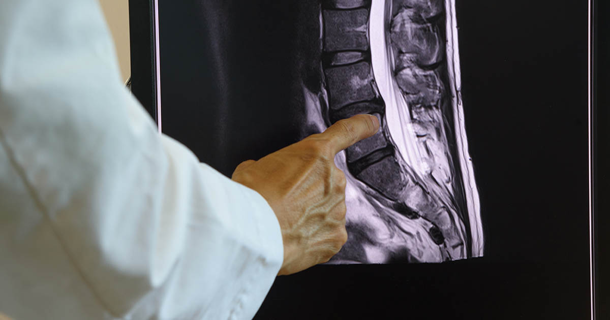
Imaging findings. A lumbar MRI can show a disc herniation clearly. It cannot reliably show SI joint dysfunction. The cartilage, ligaments, and joint space changes that drive SI joint pain are subtle on MRI, and many patients with significant SI joint dysfunction have lumbar MRI reports that look unremarkable. This is one of the most common reasons SI joint pain gets missed: the patient gets an MRI, the disc "looks fine," and everyone assumes the pain must be muscular or behavioral.
A normal lumbar MRI does not mean there is nothing wrong. The sacroiliac joint is not the focus of a standard lumbar MRI, and the joint-space changes that drive SI joint pain are often not captured. Clinical examination matters more here than imaging.
Neurological symptoms. A disc herniation that compresses a nerve root produces dermatomal numbness, tingling, and weakness that follows a predictable anatomical map down the leg. SI joint pain can refer into the leg, but it tends to produce a vaguer, heavier aching pattern rather than the sharp, electric, or burning quality of true nerve root compression. Reflexes and strength usually remain intact with SI joint dysfunction.
SI Joint Pain vs Sciatica: Overlapping Symptoms, Different Drivers
The overlap with sciatica is where patients get most confused, and understandably so. Both conditions cause pain that starts in the lower back and buttock and can radiate down the back of the leg. The key distinctions:
True sciatica is a symptom, not a diagnosis. It describes nerve root irritation, usually from a disc herniation or stenosis at L4-5 or L5-S1. The sciatic nerve carries fibers from multiple lumbar and sacral nerve roots. When one of those roots is compressed, the pain follows a dermatomal path, typically below the knee and into the foot, with associated tingling, numbness, or weakness in a specific pattern. You can read more about the disc-driven version in our post on 5 signs sciatica is disc-driven.
SI joint-referred pain typically does not go below the knee. It stays in the buttock, the posterior hip, the back of the thigh, and sometimes the groin. The neurological examination is usually normal: no weakness, no reflex changes, no dermatome-specific numbness. The pain is position-dependent rather than nerve-provoked.
One useful bedside test: the FABER test (Flexion, ABduction, External Rotation of the hip). When this movement produces the patient's familiar pain deep in the SI joint region, it suggests the SI joint or hip as the pain generator rather than the disc or nerve root. Clinicians also use the distraction test, the compression test, and the thigh thrust test. No single test is definitive, but when three or more of these provocation tests are positive, the clinical evidence for SI joint involvement is strong.
SI Joint Pain vs Piriformis Syndrome: A Common Mix-Up
There is a third condition in this neighborhood that causes similar buttock and leg symptoms: piriformis syndrome. The piriformis is a deep external rotator of the hip that sits close to the sciatic nerve. When it becomes tight or inflamed, it can irritate the sciatic nerve and produce buttock and posterior leg pain that is almost indistinguishable from SI joint dysfunction or true sciatica by symptom pattern alone.
The distinguishing feature is that piriformis syndrome is typically reproduced by hip internal rotation (turning the foot inward, which stretches the piriformis) and is often associated with a tender spot deep in the buttock rather than at the PSIS. The pain pattern from piriformis syndrome also tends to run more laterally through the buttock and hip, while SI joint pain more often involves that inner sacral region.
In practice, SI joint dysfunction and piriformis syndrome frequently coexist. The asymmetrical pelvic loading that causes one often creates compensatory tension in the other. Treatment addresses both.
Who Gets SI Joint Dysfunction and What Makes It Worse
SI joint dysfunction is more common in certain populations and with certain histories:
- Pregnancy and postpartum: Relaxin-mediated ligament laxity during pregnancy, combined with the altered gait and load distribution of carrying a baby, makes the SI joint vulnerable. Postpartum SI joint pain is significantly underdiagnosed.
- Prior lumbar surgery (fusion): When lumbar vertebrae are fused, the adjacent joints compensate by taking on more movement and load. The SI joint, just below the fusion, is a common site for this adjacent-segment stress. This is one reason many people who have had back surgery continue to have lower back pain.
- Leg length discrepancy: Even a half-inch of true leg length difference creates an asymmetrical tilt through the pelvis with every step. Over time, this asymmetry loads one SI joint more than the other.
- One-sided repetitive activity: Long-distance runners who always run the same direction on a cambered road, golfers with an asymmetrical swing, or anyone whose work involves repetitive one-sided loading (carrying a bag on one shoulder for years, for example) can develop asymmetrical SI joint stress.
- Falls and direct trauma: A fall directly onto the buttock, or a shearing force like a rear-end collision, can disrupt the normal mechanics of the SI joint acutely. This is worth noting for auto-injury patients who sometimes develop SI joint symptoms in the weeks following a crash, even when the lumbar MRI looks clean.
How We Evaluate and Treat SI Joint Dysfunction
A thorough clinical evaluation is the foundation. The history matters: when did it start, what makes it better, what makes it worse, does it radiate below the knee, are there neurological symptoms, what is the sleep position, has it happened before? The physical examination uses a battery of SI joint provocation tests alongside standard lumbar, hip, and neurological testing to build a clear picture of where the pain is actually originating.
When the clinical picture points to the SI joint, the treatment approach is different from disc-driven care:
Chiropractic mobilization of the SI joint. The SI joint responds well to specific low-force mobilization and chiropractic adjustment techniques aimed at restoring normal joint mechanics. The goal is not to "crack" the joint for its own sake but to restore the symmetrical glide and tilt that the joint is supposed to have. Most patients feel a noticeable shift in their pain pattern after the first few sessions.
Soft tissue work on the surrounding musculature. The ligaments and muscles around the SI joint, including the piriformis, the gluteus medius, and the quadratus lumborum, often develop compensatory tension and trigger points. Releasing these structures reduces the secondary pain contribution significantly.
Electrical muscle stimulation (EMS). For patients whose SI joint pain involves significant surrounding muscle guarding, EMS helps reduce the spasm pattern that keeps the joint loaded asymmetrically.
SI joint belt during the acute phase. A properly fitted sacroiliac belt compresses the iliac wings together, reducing the shear forces across the joint. It is not a long-term solution but is often useful in the acute phase to allow movement without provocation.
Stabilization exercises. Because hypermobility is a driver in many cases, specific deep stabilizer exercises targeting the transverse abdominis, multifidus, and gluteus medius are important for the long term. These are different from general core exercises; the goal is targeted co-contraction of the muscles that control pelvic stability.
For cases where conservative care does not produce adequate improvement, we discuss the full picture with the patient. In rare cases, SI joint dysfunction that is refractory to conservative care may warrant a diagnostic injection to confirm the diagnosis definitively. We coordinate with the appropriate specialist when that step is indicated.
When to Take SI Joint Pain Seriously
Most SI joint dysfunction is a mechanical problem with a mechanical solution. But certain signs warrant prompt evaluation for something more serious:
- Significant morning stiffness lasting more than 45 minutes that improves with movement (possible inflammatory arthropathy like ankylosing spondylitis, particularly in younger patients)
- Pain in both SI joints simultaneously
- Unexplained weight loss alongside SI region pain
- Bladder or bowel changes
- Night pain severe enough to wake you up and that does not ease with position change
None of these are common, but each is a reason to evaluate beyond a purely mechanical explanation. We screen for red flags as part of every new patient intake.
If you are dealing with lower back and buttock pain that does not fit the classic disc or sciatica pattern, or if you have had imaging that came back "normal" but the pain has not resolved, it is worth having the SI joint evaluated specifically. You can also learn more about the broader picture of hip pain that mimics sciatica and how these conditions overlap in practice, and for a related perspective on lower back mechanics, our post on lower back stiffness when getting out of a chair covers the facet joint side of the equation.