The MRI report says "herniated disc at L4-L5" or "disc protrusion at C5-C6." Your primary care doctor hands you a referral to a spine surgeon. That sequence feels like a verdict. For most patients, it is not. In our Lakewood Ranch office, roughly 80 to 85 percent of the herniated disc cases we see respond well to a structured course of non-surgical herniated disc treatment without ever needing to talk to a surgeon. The remaining 15 to 20 percent do need a surgical evaluation, and we refer those cases promptly. The first job is figuring out which group you are in.
That answer rarely comes from the MRI report alone. It comes from a proper clinical exam: what movements reproduce or relieve your symptoms, whether neurological signs are present, and how long the symptoms have been active. What follows is a plain-language explanation of what conservative care targets, which tools we use and why, and what the research actually shows about timelines and outcomes.
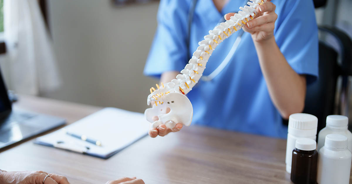
What a herniated disc is actually doing to you
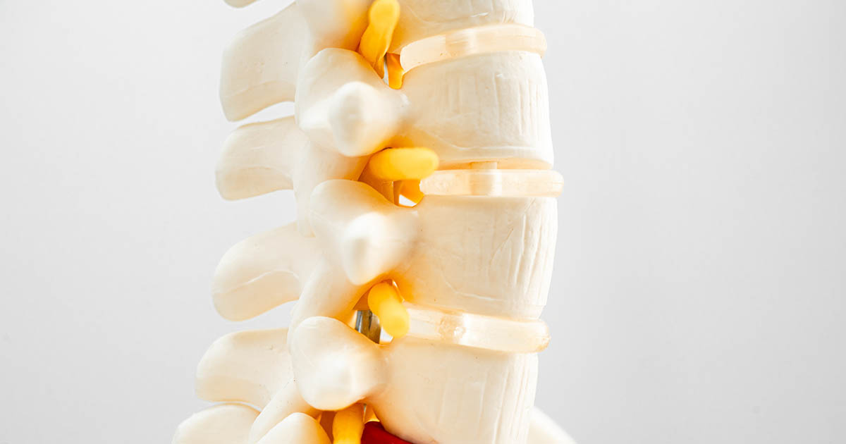
The intervertebral disc sits between two vertebrae and acts as both a shock absorber and a pivot point. The outer ring, called the annulus fibrosus, is a tough layered structure. The inner material, the nucleus pulposus, is mostly water, accounting for roughly 88 percent of the nucleus volume in healthy discs. When the annulus tears or weakens, that inner material can press outward, into the spinal canal or into the foramen where nerve roots exit.
Pain from a herniated disc has two sources. The first is chemical: the nucleus pulposus contains inflammatory compounds (phospholipase A2 is one of the better-studied ones) that irritate nerve tissue on contact. The second is mechanical: physical pressure on a nerve root alters conduction and produces the characteristic traveling pain, tingling, or weakness in the arm or leg that follows that nerve's distribution.
That distinction matters for treatment. Chemical irritation responds to approaches that reduce inflammation and promote tissue healing. Mechanical compression responds to approaches that unload the disc and create space for retraction. The best conservative programs address both.
Why most herniated discs don't end in surgery
Disc material is not a permanent fixture in the canal. Research consistently shows that herniated nucleus pulposus can and does resorb over time, particularly with larger herniations, because immune cells (macrophages) migrate to the site and gradually break down the displaced nuclear material. A landmark 1996 study in the New England Journal of Medicine found that about 90 percent of patients with herniated lumbar discs had significant improvement within 12 weeks of conservative care. Subsequent imaging studies have confirmed visible reduction of disc herniations in a substantial portion of cases after months of non-surgical management.
That natural history is worth knowing, but it cuts both ways. Waiting without doing anything often means 12 to 16 weeks of escalating pain, nerve sensitization, and progressive disc degeneration. The goal of conservative care is not to wait and see. The goal is to create the mechanical and biochemical conditions that accelerate the resorption and recovery process while keeping the nervous system functional during it.
A herniated disc can shrink. The right environment speeds that process up. The wrong environment (continued loading, inflammation, nerve compression left untreated) slows it down or reverses it.
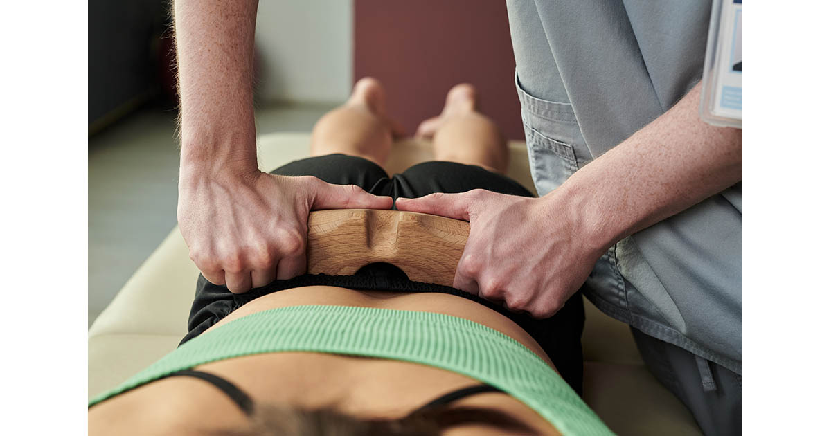
The core of conservative care: spinal decompression
If there is one tool that changes outcomes for disc herniations, it is computer-controlled non-surgical spinal decompression. This is not the same as traction, stretching, or an inversion table. Motorized decompression uses a precise, controlled distraction force applied at specific angles and intensities, guided by a computer program that adjusts in real time based on the patient's muscle guarding response.
The mechanism is specific. By creating a negative intradiscal pressure (research using intradiscal pressure transducers documents values as low as negative 150 mmHg during therapeutic decompression), the treatment does three things simultaneously:
- Draws displaced nuclear material back toward center by hydrostatic pressure gradient
- Opens the foramen where the compressed nerve root exits, reducing direct mechanical pressure
- Promotes imbibition: the disc's nutrient uptake mechanism, which is essentially a pumping action that pulls fluid and nutrients back into the dehydrated nucleus
For lumbar herniations at L4-L5 or L5-S1 (the most common locations), a full decompression protocol typically runs 20 to 24 sessions over 6 to 8 weeks. Cervical herniations at C5-C6 or C6-C7 follow a similar pattern on a different table configuration. In our experience over 23 years, patients with contained disc herniations and 8 weeks or fewer of symptoms tend to respond faster than those who waited longer. That is not a coincidence; it reflects the inflammatory timeline of disc tissue.
Supporting the disc: what we layer in and why
Decompression moves the mechanical driver. But the chemical driver (inflammation, nerve sensitization, tissue hypoxia around damaged disc tissue) requires additional tools. Here is what we use and what each one targets.
Class IV laser therapy
Class IV lasers deliver photonic energy into tissue at therapeutic depths that cold (Class II/III) lasers cannot reach. In the lumbar spine, that means penetrating through paraspinal muscle to reach the posterior disc and facet structures. The photobiomodulation effect works by stimulating mitochondrial activity (specifically cytochrome c oxidase), which increases cellular ATP production and down-regulates inflammatory prostaglandins.
In practical terms, patients report reduced pain and improved range of motion within the first several sessions. For disc herniations complicated by facet inflammation or paraspinal guarding, laser is one of the most reliable tools for breaking that secondary pain cycle. We use it routinely as part of disc herniation protocols.
Hyperbaric oxygen therapy (HBOT)
The intervertebral disc is the largest avascular structure in the human body. It has no direct blood supply; nutrition arrives by diffusion through the vertebral end plates. When disc tissue is damaged and inflamed, that diffusion environment becomes hostile to healing, particularly in the dehydrated, oxygen-deprived nucleus.
Hyperbaric oxygen dissolves oxygen directly into plasma at pressures above atmospheric, bypassing the hemoglobin carrier system. This saturates tissue beds that are otherwise poorly perfused, including the tissue surrounding a herniated disc. For patients with disc injuries complicated by persistent nerve pain or slow progress with decompression alone, HBOT can meaningfully accelerate the inflammatory resolution phase. We discuss this option explicitly with patients whose recovery has stalled after the first 4 to 6 weeks of decompression.
Electrical muscle stimulation
A herniated disc that compresses a nerve root long enough causes the muscles that nerve controls to become inhibited. You can feel this: the affected leg (or arm, for cervical cases) feels weaker, less coordinated, fatigues faster. That inhibition is not simple weakness; it is a protective neuromuscular shutdown that persists even after the disc pressure is reduced, because the motor pathways have been altered by chronic compression.
Electrical muscle stimulation (EMS) re-establishes normal motor recruitment patterns by delivering therapeutic electrical current directly to the affected muscle group. It does not replace exercise, but it restores the baseline neuromuscular activation that makes exercise and rehabilitation productive. We sequence it after the first few weeks of decompression, once the acute compression phase has been addressed.
What the realistic timeline looks like
Three weeks is not enough time to evaluate a disc herniation program. Six weeks is the minimum meaningful checkpoint. Here is what we typically see in a well-constructed protocol:
- Weeks 1 to 2: Acute pain often increases initially as the disc begins to respond. This is normal; many patients report a 20 to 30 percent increase in discomfort during the first four to six sessions before improvement begins. Setting this expectation prevents early dropout.
- Weeks 3 to 5: Most patients report their first meaningful pain reduction during this window. Range of motion improves. The radiating leg or arm symptoms (if present) often diminish before central back pain does, which reflects nerve decompression ahead of full disc resorption.
- Weeks 6 to 8: Functional improvement becomes measurable. Patients can sit longer, sleep better, and resume activities they avoided. We reassess at this point and determine whether to continue, modify, or discharge.
- Weeks 9 to 12: For cases that are progressing but not yet resolved, this phase focuses on stabilization: restoring deep lumbar stabilizer function (multifidus, transversus abdominis) to prevent recurrence. A disc that resorbs but has no muscular support around it often reherniates within 12 to 18 months.
Patients in Lakewood Ranch and Bradenton who start within 8 weeks of onset and have contained herniations typically move through this timeline faster. Those with longer symptom duration, larger herniations, or multi-level involvement take longer and require more sessions. Both outcomes are normal; the key is establishing a clear benchmark early so neither patient nor provider is operating in the dark.
When we refer out for surgery
Conservative care is appropriate for the majority of herniated disc cases. It is not appropriate for all of them, and being clear about that distinction is part of what makes the evaluation worth having.
We refer directly to a spine surgeon when any of the following are present:
- Progressive motor weakness (strength going from 4/5 to 3/5 or lower over days or weeks)
- Cauda equina syndrome: loss of bowel or bladder control, saddle anesthesia, bilateral leg weakness. This is a surgical emergency; go to the ER.
- Significant neurological deficit that has not improved after 6 to 8 weeks of appropriate conservative care
- Imaging showing severe canal stenosis (less than 50 percent of normal canal diameter) or free sequestered fragment with major nerve root compression
- Fracture, tumor, or infection as the underlying cause
The goal of conservative care is to be the right choice for the people it is right for, not to delay necessary surgery. In our experience, the cases that need surgery are usually apparent by week 6 to 8 of an honest care trial. Anything that looks like a deteriorating neurological exam before that point gets referred sooner.
What we actually do at the first visit
When a patient comes in with a herniated disc, we do not start treatment on the first appointment. We do an intake exam. That means a seated and standing postural analysis, range of motion testing in all planes, orthopedic and neurological provocation tests (straight leg raise, slump test, Spurling's for cervical), dermatomal and myotomal testing to map the nerve distribution, and a review of any existing imaging.
If you have an MRI from the last 6 months, bring it; we read it directly rather than waiting for a radiology report. If you do not have imaging and the clinical exam warrants it, we can coordinate that referral. The point of the intake exam is to build a clinical picture that drives a specific care plan, not a generic protocol.
For a deeper breakdown of what a first visit looks like, see our step-by-step guide to your first chiropractic visit. For information on herniated disc symptoms that trace back to nerve root compression, the disc-driven sciatica post covers how to distinguish the disc pattern from other sciatic causes.
If lower back pain has been your daily reality for months, the most useful step is finding out what is actually driving it. That answer shapes everything downstream. Call (727) 213-2982 or book online; we have same-week availability for new herniated disc cases.