A patient came in last month who had been dealing with a burning ache down the back of his left leg for three months. His primary care doctor ordered an MRI, the report came back saying "L5-S1 disc herniation with left S1 nerve root compression," and the discharge note said "lumbar radiculopathy." He sat across from me holding the paper, not sure what any of it meant, only knowing that the leg hurt and Tylenol was not helping.
That is a very common situation. "Lumbar radiculopathy" is one of the most frequent diagnoses in spine care, and it is also one of the least explained. Patients hear "radiculopathy" and often assume it is the same as sciatica, or a pulled muscle, or something that will go away on its own with enough rest. Sometimes one of those is true. Often, none of them are. Understanding what the diagnosis means changes what you do about it.
If you have been told you have lumbar radiculopathy and you are trying to figure out what to do next, start here: our sciatica and nerve root pain page covers how we evaluate and treat this at Spine and Wellness Center Lakewood Ranch. The rest of this post walks through the anatomy and what you need to know before your next appointment.
What "lumbar radiculopathy" actually means
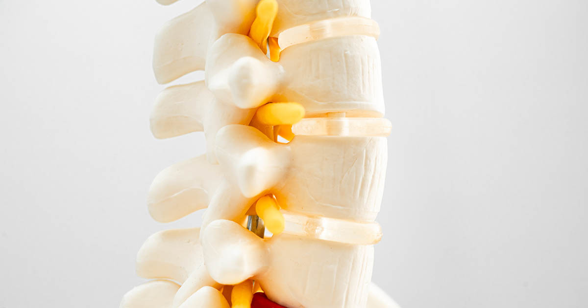
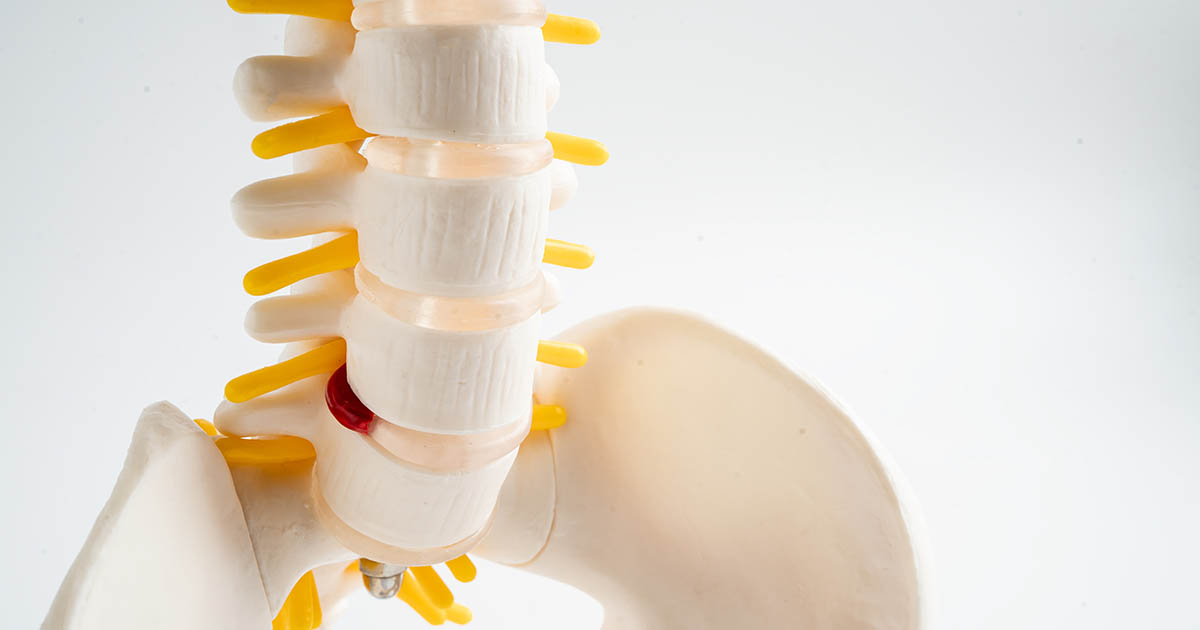
Break the word down. "Lumbar" refers to the five lowest vertebrae of the spine, L1 through L5, just above the sacrum. "Radiculopathy" comes from the Latin "radix" (root) and the Greek "pathos" (suffering or disease). A radiculopathy is a problem at the nerve root level: the point where a spinal nerve exits the spinal cord and passes through a small opening in the vertebra called the intervertebral foramen.
There are two words people use almost interchangeably that mean different things. "Sciatica" is a symptom: pain that travels down the back of the leg along the path of the sciatic nerve. "Lumbar radiculopathy" is a diagnosis: dysfunction of a specific nerve root caused by compression, inflammation, or chemical irritation. Every case of sciatica has a source somewhere. Lumbar radiculopathy is one specific source. It is usually the right answer, but not the only possibility.
What compresses a lumbar nerve root? In most patients under 50, a disc herniation is the cause. The intervertebral disc sits between two vertebrae and acts as a shock absorber. When the soft inner nucleus pushes through the tougher outer ring (the annulus), the extruded material can press directly against the nerve root that is just behind it. Bone spurs, a narrowed foramen from arthritis, and ligament thickening can cause the same compression. The result in all cases is the same: the nerve root cannot function normally, and it tells you so by producing pain, numbness, or weakness along its entire length.
Lumbar radiculopathy is not a muscle problem. The burning, the tingling, the shooting pain down the leg: those come from a nerve. Treating only the muscle will not fix the nerve root. That distinction matters for every treatment decision that follows.
Which nerve root, which symptoms: the L3 through S1 map
Each lumbar nerve root controls a predictable strip of skin (a dermatome) and a specific set of muscles. When a root is compressed, the symptoms follow its territory. This is how a clinician can often identify the level from a physical exam before even looking at an MRI. Here is a practical level-by-level breakdown.
L3 and L4 radiculopathy
L3 and L4 nerve roots exit at the upper and mid-lumbar spine. Compression here produces pain and numbness that travels from the front of the thigh down toward the inner knee and the inside of the lower leg. Weakness shows up in the quadriceps: the muscle that straightens the knee. A key reflex test is the patellar (knee jerk) reflex, which is reduced or absent when L4 is involved. People with L3 or L4 radiculopathy often notice that stairs and squatting become harder, and there may be a burning or aching sensation on the anterior thigh that does not quite match typical sciatica descriptions. L3-L4 disc herniations account for roughly 10 percent of lumbar disc problems but tend to produce more pronounced quad weakness than lower-level lesions.
L5 radiculopathy
L5 is the single most commonly involved nerve root in lumbar disc herniation, usually from an L4-L5 disc. The L5 dermatome covers the outer (lateral) lower leg, the top of the foot, and the big toe. Weakness shows in the ability to lift the foot upward: dorsiflexion. A patient with significant L5 involvement may notice their foot drops or drags slightly when walking, particularly when they are fatigued. There is often a deep ache in the outer buttock that radiates down the outer shin to the top of the foot. The big toe becomes hard to extend against resistance. The Achilles reflex is usually intact at this level; it is the L4 patellar reflex and S1 Achilles that test differently.
S1 radiculopathy
S1 is the other very common root involved in lower lumbar disc herniations, typically at L5-S1. The S1 dermatome covers the back of the calf, the heel, and the outer edge of the foot (the small-toe side). Pain and numbness in this distribution is what most people think of when they picture "sciatica": a shooting sensation down the posterior (back) of the leg into the foot. Weakness shows up in calf raise ability, and the Achilles reflex is reduced or absent. Many patients notice they cannot stand on their tiptoes as long on the affected side. The pain tends to worsen sitting, especially with the leg extended, and eases somewhat when standing or walking slowly.
Identifying which level is involved matters because the disc at that level is the treatment target, and the weakness patterns tell you how far along the nerve dysfunction is. A patient with measurable quad weakness from L4 compression may need a faster treatment response than someone with only sensory changes.
How this is different from other causes of leg pain
Leg pain that follows a nerve pattern is not the same as leg pain from a muscle strain or a joint problem. A few features help distinguish radiculopathy from other causes:
- Dermatomal distribution. The numbness or tingling follows a specific nerve map, often down to the level of which toes are affected. Muscle strains produce diffuse achiness without this specificity.
- Neurological signs. Reflex changes, weakness in a specific muscle group, and sensory changes on nerve testing point toward a nerve root, not a muscle.
- Positive straight leg raise. In this clinical test, the patient lies flat and the leg is raised with the knee straight. At a certain angle, this stretches the nerve root. If a sharp pain or electrical sensation shoots down the leg below the knee at 30 to 70 degrees, the test is considered positive and strongly suggests L4, L5, or S1 involvement. A negative SLR does not rule out radiculopathy but does change the picture.
- Position dependence. Disc-driven radiculopathy typically worsens with sitting (which increases disc pressure), with bending forward, and with the Valsalva maneuver (coughing, sneezing). It tends to ease with walking slowly or lying down in certain positions. Vascular causes of leg pain, by contrast, worsen with walking and ease with rest.
Piriformis syndrome is frequently confused with lumbar radiculopathy because it also produces buttock pain with sciatic nerve involvement. The key difference: piriformis syndrome is peripheral compression (the piriformis muscle compressing the sciatic nerve in the hip), not a spinal level issue. Treatment is different and so is the source. Our post on pinched nerve pain covers how we differentiate between spinal and extra-spinal nerve compression during evaluation.
Does every lumbar radiculopathy need an MRI right away?
Not immediately, but it depends on what is happening neurologically. This is a common question and the answer requires nuance.
For most patients with a new lumbar radiculopathy and primarily sensory symptoms (pain and numbness without significant weakness), a 4 to 6 week trial of active conservative care is reasonable before MRI. The clinical reality is that a large percentage of lumbar disc herniations improve or resolve through a combination of anti-inflammatory response, careful loading of the disc, and specific movement patterns that encourage the disc material to retract.
MRI moves forward immediately when:
- Rapid or progressive motor weakness (difficulty lifting the foot, buckling knee, new limping)
- Bilateral symptoms (both legs affected)
- Bladder or bowel changes (this is a medical emergency called cauda equina syndrome, requiring same-day evaluation)
- Symptoms following recent significant trauma
- Known cancer history with new spine pain
- Fever with back and leg pain (possible infection)
- Failure to improve after 6 to 8 weeks of structured conservative care
At Spine and Wellness Center Lakewood Ranch, Dr. Banman screens for all of these red flags at the first evaluation. If any are present, you will be referred immediately to the appropriate specialist or imaging center. The goal is never to delay necessary care. The goal is also not to send every patient with leg pain to MRI on day one, because early imaging often shows disc changes that are not actually causing the current symptoms, and can lead to unnecessary anxiety or surgical conversations before conservative care has had a chance to work.
What conservative care can realistically accomplish
The good news: most lumbar radiculopathies, even those with clear nerve root compression on imaging, respond to conservative care when that care is structured and specific. The key concept is centralization. Centralization is when pain that was spreading down the leg begins to retreat back toward the lower back during treatment. It is a reliable clinical sign that the disc herniation is reducing its pressure on the nerve root. Not every patient centralizes, but many do, and those who do reliably report better outcomes.
At our clinic, the core tools for lumbar radiculopathy include:
- Non-surgical spinal decompression. The DOC-20 table creates a specific distraction force at the targeted disc level, reducing intradiscal pressure and allowing the nucleus to retract. This is very different from general traction. The computer-controlled distraction prevents the paraspinal muscles from splinting, which is what limits manual traction. For L4-L5 and L5-S1 herniations producing radiculopathy, spinal decompression is often the anchor of the care plan.
- Class IV laser therapy. The nerve root in lumbar radiculopathy is not just mechanically compressed; it is also chemically inflamed by substances released from the disc. Class IV laser uses deep tissue photobiomodulation to reduce that periradicular inflammation, which can accelerate the reduction in leg pain even when the disc is still pressing on the root.
- Chiropractic adjustments. Targeted at the specific facet joints around the involved level, not generalized cracking. Joint mobility at the affected segment affects the size of the intervertebral foramen. A restricted facet joint that narrows the foramen adds to the compression.
- Neurological exercises. McKenzie-based extension loading, when the patient centralizes with extension, is a significant part of care. Patients learn which positions load the nerve root and which positions decompress it, and build that into their daily habits.
For patients dealing with ongoing nerve inflammation from a long-standing herniation, our neuropathy tools (ReBuilder therapy, EMS, whole-body vibration for neuromuscular activation) can support nerve fiber function during the recovery period. This is especially relevant when there is measurable motor weakness or significant sensory loss. We cover the full multi-tool approach in our back and nerve pain care overview.
Timeline: what to expect and when to reassess
Many patients with acute lumbar radiculopathy (symptoms under six weeks) see meaningful pain reduction within three to six weeks of structured conservative care. The leg pain tends to decrease and centralize before the back pain fully resolves. That sequence is actually a good sign.
Chronic radiculopathy (symptoms over three months) takes longer, often three to six months of consistent care. When there is established motor weakness, recovery of the muscle function may lag behind pain relief by weeks to months, because nerve fibers regrow slowly. Progress happens but the timeline requires patience.
The situations where conservative care typically cannot produce adequate improvement include: very large disc herniations that have sequestered (broken free) and are directly impacting the nerve; severe progressive weakness; and cases where the compression is primarily from bone rather than disc material, particularly in older patients with multilevel stenosis. In those cases, referral to a spine surgeon for an honest surgical conversation is the right next step. Dr. Banman has those conversations directly with patients and does not carry a financial incentive to push conservative care indefinitely when it is not working.
If you are in Lakewood Ranch, Bradenton, or Sarasota and you have been told you have lumbar radiculopathy, a structured evaluation will tell you within the first visit which nerve root is involved, which level the disc problem is at, whether centralization is possible with your specific pattern, and what a realistic care plan and timeline look like. That is a very different starting point than "take ibuprofen and wait."
Common questions patients ask at the first visit
A few things come up in almost every lumbar radiculopathy evaluation, so here is how Dr. Banman typically answers them.
Will this go away on its own? Some do. A portion of disc herniations, particularly soft nuclear herniations, undergo natural resorption over months. But "waiting it out" without structured care tends to produce slower results, and it gives the nerve more time under compression, which increases the risk of lasting weakness or sensory change. Doing nothing is a choice, but not usually the best one.
Should I stop moving? No. Complete rest is counterproductive for most lumbar radiculopathies. The disc needs movement to hydrate and the nerve root benefits from gentle motion. The goal is to identify which specific movements aggravate versus relieve the nerve root and build your activity around that pattern.
Is surgery the next step? For most acute radiculopathies, surgery is not the next step. It is an option when conservative care has been given a genuine structured trial (typically six to eight weeks minimum) and there is either insufficient improvement or progressive neurological deterioration. Most lumbar microdiscectomies are elective, not emergency procedures, and they perform similarly to conservative care in the majority of patients at one to two years out. The surgical advantage tends to be faster short-term pain relief. The conservative advantage is avoiding the risks and recovery of surgery. That tradeoff is worth understanding before any decision is made.
Can I still exercise? Almost always yes, with modifications. Walking, swimming, and specific core exercises that do not flex or load the affected disc level are usually fine and beneficial. Heavy deadlifts, intense bending, and prolonged sitting are the main things to modify during active treatment. Dr. Banman walks through which movements are safe for each patient's specific level and severity at the first visit.