You get an X-ray for lower back pain that has dragged on for months and the radiologist's report comes back with a word you have never seen before: spondylolisthesis. The report might describe it as Grade I or Grade II, at L4-L5 or L5-S1, and note whether it is stable or progressive. None of that tells you what it actually means for your day-to-day life or what you can do about it.
This post breaks down what spondylolisthesis is, why it causes the symptoms it does, what separates people who do fine from people who do not, and what a realistic conservative care plan looks like in Lakewood Ranch.
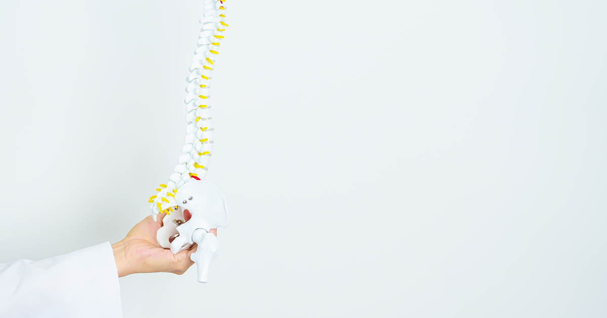
What spondylolisthesis actually is
The word comes from the Greek "spondylos" (vertebra) and "olisthesis" (slip). In plain terms, one vertebra has shifted forward relative to the vertebra directly below it. The most common site is L5 slipping forward on S1, followed by L4 slipping on L5. These two levels carry the highest mechanical loads in the entire spine, which is why slippage happens there first.
The degree of slip is classified in four grades, based on what percentage of the vertebral body has moved across the one below:
- Grade I: 1 to 25 percent slip. The most common finding. Most patients with Grade I are pain-free or have manageable symptoms with conservative care.
- Grade II: 26 to 50 percent slip. Symptoms are more common and tend to be more persistent. Conservative care still produces real results for many patients.
- Grade III: 51 to 75 percent slip. More likely to involve neurological findings. Conservative care is still attempted, but surgical consultation becomes part of the conversation.
- Grade IV: 76 to 100 percent slip. Rare. Neurological compromise is frequent. Surgical evaluation is typically indicated.
The vast majority of spondylolisthesis patients seen in a chiropractic office are Grade I or II. Grade III and IV are uncommon outside of trauma or severe developmental presentations.
Why does a vertebra slip in the first place?
There are several distinct mechanisms, and the type matters because it influences how the condition behaves and responds to care.
Isthmic spondylolisthesis (the most common type in active patients)
This involves a stress fracture or defect in a small bony bridge called the pars interarticularis, the narrow section connecting the facet joints at the back of each vertebra. When the pars fractures on both sides (a condition called spondylolysis), the vertebra loses its rear anchor and can slide forward under the mechanical load of walking, extension, and rotation.
Isthmic spondylolisthesis is most often seen in athletes and formerly athletic adults who loaded their lumbar spines heavily in extension: gymnasts, football linemen, weight lifters, dancers. It is usually identified in adolescence but may not become symptomatic until adulthood when discs begin to lose height.
Degenerative spondylolisthesis (the most common type in adults over 50)
Here the pars is intact. The slip happens because the facet joints and discs have degenerated enough to allow forward translation that they would normally resist. Think of the disc as a shock absorber and the facet joints as the rear locks on a drawer. When both wear down, the drawer can slide forward.
Degenerative spondylolisthesis is strongly associated with disc degeneration at the same level, and it is more common in women (the facet joint geometry at L4-L5 in women allows slightly more forward shear). The L4-L5 level is affected more often than L5-S1 in this type.
The most important thing to understand about degenerative spondylolisthesis: the slip itself is rarely the only problem. It is almost always accompanied by disc narrowing, facet arthritis, and some degree of central or foraminal canal narrowing at that level. The pain and nerve symptoms usually come from the combination, not from the slip in isolation.
Congenital and traumatic types
Congenital spondylolisthesis results from malformation of the posterior elements at birth. Traumatic spondylolisthesis follows a fracture that damages the facets, lamina, or pedicles. Both are less common in the general patient population and are typically identified earlier in life.
What symptoms does spondylolisthesis cause?
The symptom picture varies widely depending on the grade, the type, the level, and whether nerve structures are being compressed. Some patients with confirmed Grade I spondylolisthesis have no symptoms at all and discover it incidentally on imaging ordered for something else. Others have significant daily limitations.
The most common presentations:
- Lower back pain that worsens with standing and extension. Because extension loads the posterior structures and narrows the canal, people with spondylolisthesis often feel best when flexed slightly forward (sitting, leaning on a shopping cart) and worst when standing erect for long periods or arching backward.
- Buttock and thigh pain. Often bilateral. The L4-L5 or L5-S1 slippage can narrow the central canal and put pressure on the nerve roots that feed the buttocks and posterior thighs.
- Radiating leg pain (radiculopathy). When the slippage significantly narrows the foramen (the opening through which nerve roots exit), one or both legs may develop the classic sciatica pattern: burning, shooting, or electric-shock pain from the buttock into the leg, sometimes with numbness or weakness.
- Neurogenic claudication. This is a walking-distance limitation in which leg pain, heaviness, or cramping builds as you walk and eases when you sit or lean forward. It is the hallmark of spinal stenosis and can accompany higher-grade spondylolisthesis when there is significant central canal narrowing.
- Step-off deformity. In higher grades, you can sometimes feel or see a visible step at the lower back where the slipped vertebra creates a ledge. A physical exam finding, not typically noticeable by the patient themselves.
Red flags that indicate the need for urgent evaluation: loss of bowel or bladder control, saddle area numbness, rapidly progressing weakness in one or both legs. These suggest cauda equina involvement and require same-day emergency assessment.
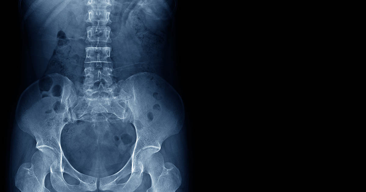
How is spondylolisthesis diagnosed?
Plain X-rays (standing, weight-bearing) are the standard first step and will usually reveal the slip, the grade, and whether it changes with flexion and extension (indicating instability). The lateral view is the key one.
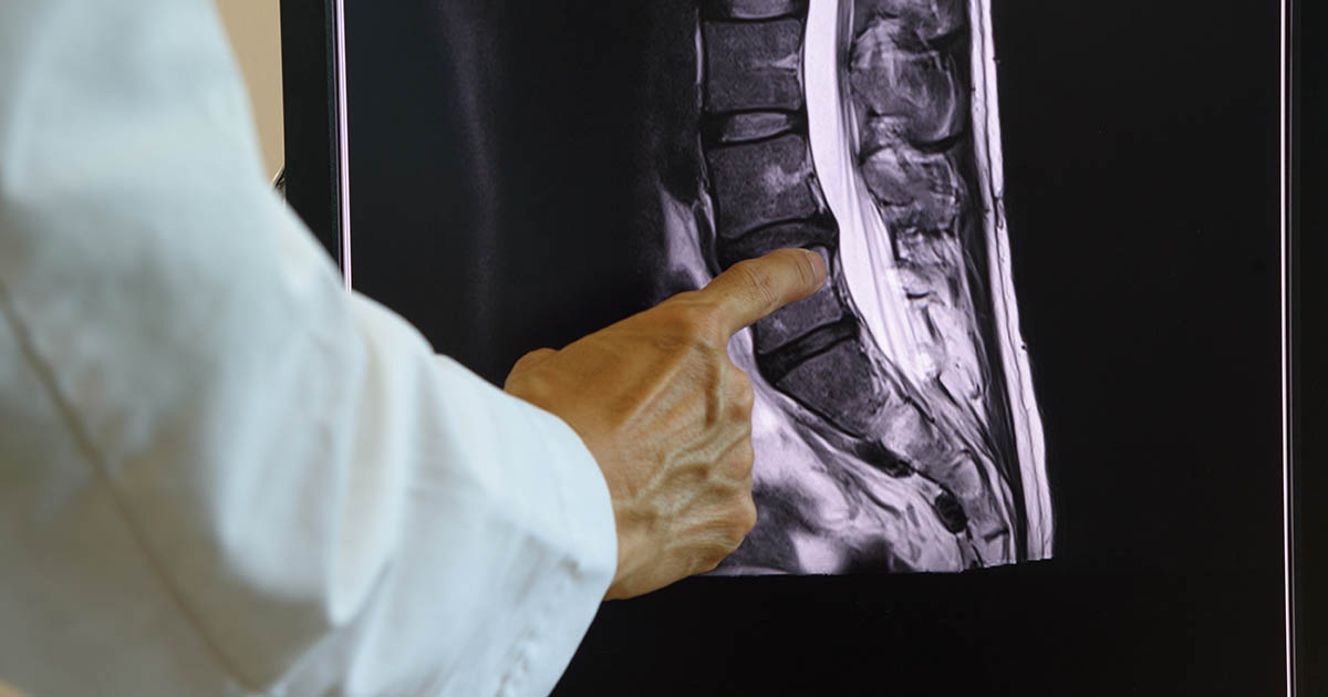
MRI adds information about the soft tissue components: disc condition at the slipped level, degree of canal and foraminal narrowing, and nerve root involvement. MRI is indicated when there are neurological symptoms (leg pain, numbness, weakness) or when conservative care is not producing the expected response after a reasonable trial.
CT scan is useful for bony detail, particularly to confirm whether a pars defect is present and whether it is acute (fresh stress fracture with reactive bone) or chronic (sclerotic, well-established).
One practical point: grade on imaging does not always predict symptom severity. Some Grade II patients function well with conservative care. Some Grade I patients have significant nerve symptoms because of accompanying foraminal stenosis. We evaluate both the imaging and the clinical presentation together, not imaging alone.
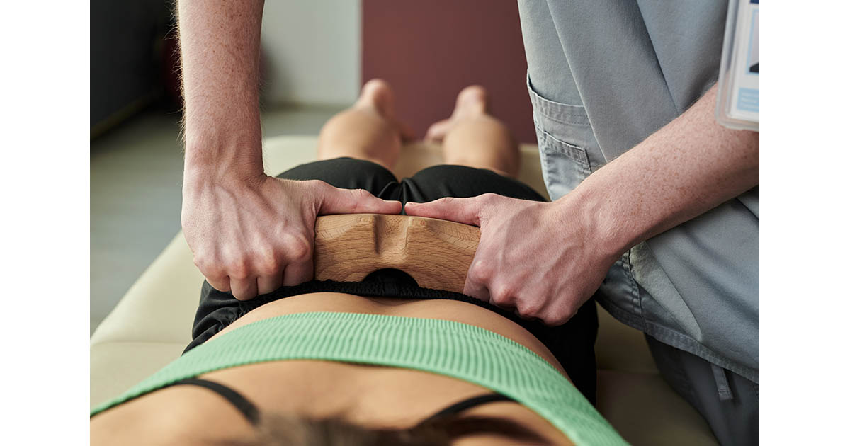
What conservative care can do
For Grade I and most Grade II spondylolisthesis, the evidence supports conservative care as the first-line approach. That does not mean rest. It means active, structured treatment aimed at stabilizing the slipped segment, reducing nerve irritation, and teaching the body to load the spine safely.
At our Lakewood Ranch clinic, the conservative approach for spondylolisthesis typically includes:
Spinal decompression
Computer-guided traction that gently unloads the affected disc and reduces pressure on the nerve roots exiting around the slipped level. For patients whose primary symptom is leg pain or neurogenic claudication, spinal decompression can produce meaningful relief by reducing foraminal compression. We select patients for decompression carefully: it is not appropriate when the slip is significantly unstable or when there is active spondylolysis that could propagate under traction.
Chiropractic care tailored to the instability
Standard high-velocity lumbar adjustments are modified or avoided at the slipped level itself. Instead, the focus is on adjacent levels that become hypermobile to compensate, on pelvic alignment, and on reducing the muscle guarding that builds up around an unstable segment. Instrument-assisted techniques and soft tissue work play a larger role here than they do in a straightforward disc herniation.
Class IV laser therapy
For the inflammatory component driving nerve irritation around the slipped level, Class IV laser delivers photonic energy that supports tissue repair and reduces local inflammation. Many patients report reduced pain intensity after a series of laser treatments, particularly when combined with decompression.
Core stabilization and movement retraining
The deeper spinal stabilizers, the multifidus and transverse abdominis, are often inhibited in people with chronic lower back pain. Rebuilding their function helps create a muscular brace around the unstable segment. This is progressive and supervised, not generic back exercises.
Whole body vibration for neuromuscular activation
Low-frequency whole body vibration stimulates the muscle spindles and deep stabilizers without compressive loading, which makes it a useful adjunct for spondylolisthesis patients who cannot yet tolerate conventional exercise loads.
What realistic outcomes look like
The research on conservative care for Grade I and II degenerative spondylolisthesis is reasonably positive. Many patients achieve meaningful pain reduction and functional improvement with a structured 8 to 12 week program. The goal is not to reverse the slip (conservative care does not reverse established spondylolisthesis) but to reduce the neural irritation driving the symptoms, stabilize the segment, and restore enough function that the condition is manageable.
Some patients do well for years with periodic maintenance care. Others plateau after initial improvement and require surgical consultation. Factors that predict a less favorable conservative response include: significant central canal stenosis causing neurogenic claudication, Grade III or IV slip, active neurological deficits that are not improving, and clear instability on flexion-extension X-rays.
If you are in that category, we will tell you directly and help coordinate a surgical referral. Surgery for spondylolisthesis typically involves fusion, and the outcomes for well-selected patients with Grade II or higher slips causing neurological symptoms are generally good. We continue to coordinate post-surgical rehabilitation for patients who have had spinal fusions.
For patients who have had prior auto accidents and notice their spondylolisthesis symptoms worsening in the weeks after a collision: this is a recognized pattern. An existing spondylolisthesis can be aggravated by the forces of a crash even at moderate speeds. We document this carefully as part of auto-injury care, and we coordinate with attorneys when relevant.
Questions patients commonly ask
Does spondylolisthesis always get worse over time? Not necessarily. Many Grade I slips remain stable for years. Grade II degenerative slips can progress, particularly if disc height at that level is lost significantly. Regular monitoring with weight-bearing X-rays (every one to two years) is reasonable for established cases.
Can I exercise? Most people with Grade I and II spondylolisthesis can exercise, with modifications. High-impact, heavy axial loading, and aggressive lumbar extension are generally avoided in the early phase. Swimming, cycling, and walking on flat terrain tend to be well-tolerated. We assess each patient individually.
Is there a brace for this? Bracing is occasionally used for acute isthmic spondylolisthesis in adolescents to reduce load during the healing phase of a pars stress fracture. For degenerative spondylolisthesis in adults, bracing is less commonly recommended for long-term use because it can lead to muscle atrophy. Short-term use during a flare-up is different.
What about injections? Epidural steroid injections can reduce the neurogenic component of the pain and may be used as part of a broader conservative program. They are not curative and do not address the mechanical instability. We coordinate with pain management physicians when injections are part of the plan.
If your imaging report uses the term spondylolisthesis and you are not sure what it means for you, we are happy to review both the report and the clinical picture together. Call us at (727) 213-2982 or book online at celluron.janeapp.com.