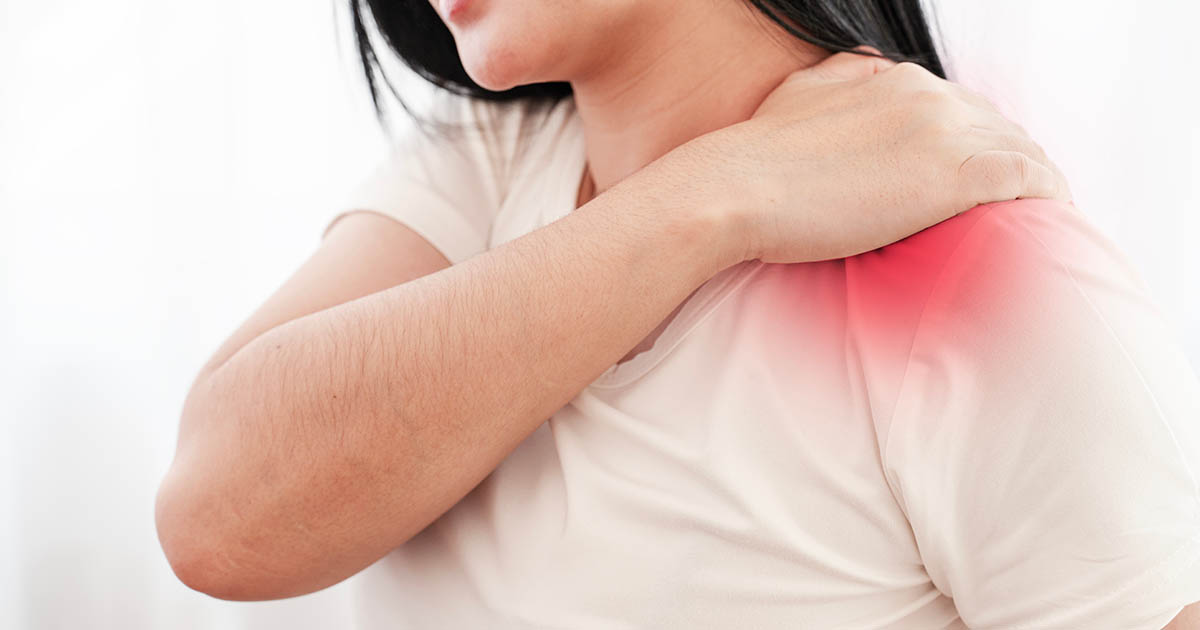
Most neck pain stays in the neck. It is annoying, it limits how far you can turn your head, and it usually responds to a few days of rest or heat. But a subset of patients comes in with something different: neck pain that travels. A burning line down the back of the arm. Tingling in specific fingers. Weakness when they try to grip a coffee mug. That presentation is almost never just a muscle problem, and treating it like one is why so many of these patients spend months going in circles.
The clinical term is cervical radiculopathy. It means a nerve root in your cervical spine is being compressed or irritated at the point where it exits the spinal canal. Our neck pain and headaches page covers the broader landscape of cervical problems; this post goes deep on the specific condition where the nerve itself is involved, because the evaluation and the treatment differ from ordinary neck pain in important ways.
After 23 years working with cervical patients at our Lakewood Ranch office, Dr. Banman estimates this presentation accounts for roughly one in four of the neck cases he sees. It is common enough to deserve a clear explanation.
What is actually happening in the spine
The cervical spine has seven vertebrae, labeled C1 through C7. Between each pair of adjacent vertebrae, a nerve root exits on each side through an opening called the foramen. These nerve roots form the basis of the brachial plexus, the network that controls sensation and motor function throughout your shoulder, arm, and hand.
When something narrows that foramen or presses directly on a nerve root, the result is radiculopathy: pain, numbness, tingling, or weakness that follows a predictable map (called a dermatome or myotome) into the arm. The location and character of symptoms tell you a great deal about which level is involved:
- C5 compression: Pain into the outer shoulder and upper arm. Weakness in shoulder abduction (lifting the arm to the side).
- C6 compression: Pain and tingling into the thumb and index finger. Weakness in wrist extension. Very common.
- C7 compression: Tingling into the middle finger. Triceps weakness. Also very common.
- C8 compression: Tingling into the ring and little finger. Grip weakness. Often confused with carpal tunnel.
Knowing this map is exactly how a thorough clinician can often tell you what level is involved before imaging confirms it. Symptoms rarely follow this map perfectly, but they rarely deviate wildly from it either.
What causes the nerve root to be compressed
Three structures are responsible for the vast majority of cases:
1. A herniated or bulging cervical disc. The discs between your cervical vertebrae are small, but they carry real load and they dehydrate with age and repetitive stress. When a disc bulges posterolaterally (backward and to the side), it can intrude directly into the foramen and press against the exiting nerve root. Disc-driven radiculopathy often has a sharper, more acute onset. Patients sometimes remember the exact moment it started, often during a forceful movement, a heavy lift, or just an awkward sleep position. For a full explanation of what happens inside the disc, see our post on herniated disc care.
2. Cervical stenosis with foraminal narrowing. As the discs thin over years and the facet joints remodel, the foramina gradually narrow. This type of radiculopathy tends to develop more slowly, affects patients in their 50s and beyond more often, and is typically provoked by extension of the neck (looking up). Many patients with this presentation report that looking down gives them some relief.
3. Uncinate process hypertrophy (bone spurs at the joints of Luschka). These small bone growths form at the lateral edges of the cervical vertebral bodies and can narrow the foramen from a different angle. Less common than the first two, but worth noting because they show clearly on imaging and affect which treatments apply.
The mistake most patients make is waiting for the arm symptoms to go away on their own. Neck pain often does resolve without intervention. But when nerve compression is the underlying mechanism, the irritation tends to persist or worsen until something physically addresses the cause. The longer a nerve root is compressed, the more likely it is that some of the sensory changes take longer to fully resolve.
How this is evaluated (and what the exam actually tells you)
Diagnosing cervical radiculopathy is primarily a clinical exercise. Imaging confirms the level and rules out serious causes, but the exam gives you the working hypothesis you need to start treatment.
Key physical examination components include:
- Spurling's test: The examiner gently compresses down through the top of the head while rotating and laterally flexing the neck toward the symptomatic side. If this reproduces arm pain, sensitivity for radiculopathy is high. It is not perfect, but a positive Spurling's in the right clinical context is meaningful.
- Upper limb tension tests (ULTT): These are neurodynamic tests that put the brachial plexus under stretch. Reproduction of arm symptoms suggests neural involvement rather than purely muscular tightness.
- Cervical distraction test: The examiner manually distracts (separates) the head from the neck. If arm pain decreases, that suggests compression is driving the symptoms and decompressive approaches are more likely to help.
- Reflexes and myotomes: Checking biceps (C5-C6), brachioradialis (C6), and triceps (C7) reflexes, alongside grip and shoulder strength, helps map the level clinically.
At Spine and Wellness Center in Lakewood Ranch, Dr. Banman runs this full clinical picture before any imaging discussion happens. Many patients arrive having already seen someone who ordered an MRI without doing a thorough neurological exam. The MRI result can be misleading without the clinical context: disc bulges are extremely common in adults over 40, and many of them are not the source of the symptoms the patient is experiencing.
When imaging is appropriate and what to expect
Imaging is indicated when the clinical picture is unclear, when symptoms are severe or rapidly worsening, when conservative care over 4 to 6 weeks has not produced improvement, or when there are neurological red flags (significant weakness, loss of reflexes, signs of myelopathy).
MRI is the modality of choice for soft-tissue detail: disc height, disc hydration, nerve root appearance, and spinal cord status. If MRI is contraindicated (pacemaker, certain implants), a CT myelogram provides comparable detail of the bony foramina.
Plain X-rays are useful for assessing disc height loss and foraminal narrowing at multiple levels but do not show disc tissue or nerve roots directly. They are often the first step in an imaging sequence.
One thing worth understanding: imaging alone does not determine treatment. A large disc herniation on MRI in a patient whose arm symptoms are improving with care is often managed conservatively. A smaller herniation in a patient with rapidly progressing weakness may warrant earlier specialist consultation. The clinical picture drives the decision, not the size of the finding on the scan.
What non-surgical treatment actually involves
The majority of cervical radiculopathy cases, particularly disc-driven ones in patients under 60, improve with structured conservative care over 6 to 12 weeks. "Conservative care" is not a euphemism for doing nothing; in this context it means a coordinated set of interventions aimed at reducing nerve root compression and irritation.
At our Lakewood Ranch clinic, the approach for cervical radiculopathy typically involves some combination of the following:
Cervical traction and decompression. Mechanical or instrument-assisted traction separates the cervical vertebrae, temporarily widening the foramina. For disc-driven radiculopathy, this is often the most directly relevant intervention because it reduces the compressive load on the affected nerve root. Many patients report that arm symptoms decrease during traction, which is a useful clinical signal. Our spinal decompression page explains how the technology works in more detail.
Chiropractic manipulation (where appropriate). Cervical manipulation is approached carefully and selectively in radiculopathy cases. When indicated, specific low-force adjustments aimed at restoring segmental mobility can reduce the inflammatory response and improve nerve root mobility. When a herniated disc is acutely inflamed, the approach is modified accordingly.
Class IV laser therapy. At the nerve root level, laser therapy at therapeutic doses promotes local circulation, reduces edema around the nerve, and supports tissue repair. It is not a replacement for mechanical decompression in true compression cases, but it reduces the inflammatory component that amplifies symptoms.
Therapeutic exercise and posture correction. Cervical stabilization work and scapular strengthening reduce the ongoing mechanical load on the cervical segments. Many cervical radiculopathy patients have forward head posture that increases foraminal compressive forces, and addressing this is part of a durable resolution rather than just symptom management.
EMS (electrical muscle stimulation) for paraspinal inhibition. In acute phases, paraspinal muscle guarding can hold the cervical spine in a position that increases foraminal compression. EMS to the cervical paraspinals addresses that muscular component.
Red flags that change the picture
Most cervical radiculopathy cases are manageable outside of a surgical setting. But a subset of presentations requires urgent specialist evaluation. These are the signals we watch for:
- Rapidly progressing arm weakness (not just pain, but actual loss of strength) over days
- Loss of bowel or bladder control (this suggests cervical myelopathy affecting the spinal cord, not just a root)
- Bilateral arm symptoms (both sides simultaneously)
- Severe constant pain unresponsive to any position change
- Symptoms following trauma, especially with mechanism of injury suggesting possible instability
If any of these are present, the correct step is a prompt referral to a neurosurgeon or spine-specialized orthopedist for evaluation, not weeks of conservative management. We coordinate this referral directly and will send your clinical documentation and imaging with you.
What the typical timeline looks like
Patients with disc-driven cervical radiculopathy in the 35 to 55 age range, presenting within a few weeks of symptom onset, tend to do well with 8 to 12 weeks of structured care. Arm tingling often improves before neck pain fully resolves; that sequencing makes sense anatomically.
Patients with stenotic foraminal narrowing and older-onset radiculopathy respond more slowly and tend to need longer maintenance intervals to stay well. "Resolved" in that context often means "well-managed" rather than "structurally corrected," because the bony changes are permanent. The goal in that group is keeping the soft-tissue component under control and maintaining enough foraminal space through posture and segmental mobility work.
Either way, the pattern we see most often in our Lakewood Ranch office is patients who waited too long, tried stretching and over-the-counter medication for several months, and arrived with a more established pattern of nerve sensitization that responds more gradually. Starting earlier, when the disc is still acutely herniated but before secondary sensitization sets in, is generally the faster path.
How to get an accurate picture of what is driving your arm pain
If your neck pain has been joined by any consistent arm, hand, or finger symptoms, the most useful thing you can do is get a thorough clinical evaluation that maps those symptoms to a spinal level. That evaluation takes about an hour and tells you whether what you have is a disc problem, a stenotic problem, a soft-tissue referral (not actually radiculopathy), or something that warrants imaging before you start any treatment.
Dr. Banman sees new cervical patients at our Lakewood Ranch office with same-week scheduling most weeks. If you are dealing with neck pain and arm symptoms in the Bradenton or Sarasota area and want a clear read on what is happening, call (727) 213-2982 or book directly at the link below.
For more context on what we see in the neck and how we approach it, visit our neck pain and headaches page or read about how to tell a pinched nerve in the neck apart from one in the shoulder, a common diagnostic confusion in this category of patients.