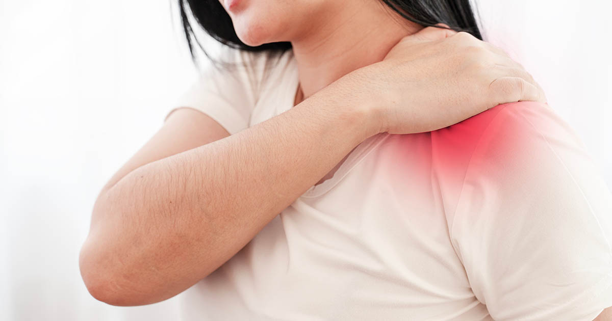
A patient came in recently describing pain that started in her neck, radiated toward her shoulder blade, and occasionally ran a burning line down her upper arm toward her elbow. She had been treating it as a shoulder problem for six weeks: stretching the rotator cuff, icing the deltoid, avoiding overhead work. Nothing helped. When we assessed the cervical spine, the pattern became clear: she had a C6 nerve root compression from a disc bulge at C5-C6. The shoulder itself was structurally fine.
This is not an unusual story. Cervical radiculopathy (a pinched nerve in the neck) and shoulder impingement are two of the most commonly confused pain patterns we see. The overlap in symptoms is real, and without a thorough structural exam, it is easy to treat the wrong system for months.
Here is how to distinguish them and what each one actually requires.
What a Pinched Nerve in the Neck Actually Feels Like
A pinched nerve in the neck, formally called cervical radiculopathy, occurs when a nerve root exiting the cervical spine is compressed or irritated. The most common cause is a disc herniation or bulge that encroaches on the neuroforamen (the opening through which the nerve exits). Bone spurs from degenerative disc disease can also cause the same compression, particularly in patients over 50.
The nerve roots most commonly affected are C5, C6, and C7. Each produces a distinct pattern:
- C5 compression: Pain and weakness in the lateral shoulder and upper arm. Some patients describe it as a deep ache in the deltoid that does not respond to shoulder stretching. Shoulder abduction may feel weak. No finger numbness.
- C6 compression: Pain travels from the neck down the outer forearm to the thumb and index finger. Biceps may feel weak or have reduced reflex. Tingling or numbness in the first two fingers is common.
- C7 compression: Pain runs from the neck through the triceps area to the middle finger. Grip may feel weaker. Triceps reflex is often reduced.
The defining characteristic of cervical radiculopathy is that neck movement changes the pain. Extending or rotating the neck toward the painful side typically worsens it (a positive Spurling sign). Conversely, many patients find partial relief by raising the arm overhead (the shoulder abduction relief sign), because this posture momentarily unloads the compressed nerve root.
If turning your head to one side reproduces, worsens, or causes pain to shoot down your arm, the neck is involved. That is not a shoulder symptom.
Other characteristics common in cervical radiculopathy: the pain often has a burning or electric quality, it may wake patients at night if they roll onto the affected side, and it is frequently described as running in a line rather than sitting in one spot.
What Shoulder Pain Actually Feels Like
True shoulder pathology, whether rotator cuff tendinopathy, impingement syndrome, a partial rotator cuff tear, or frozen shoulder (adhesive capsulitis), produces pain that behaves very differently.
The main distinctions:
- Location is localized. Shoulder pain tends to sit in the deltoid region, the anterior shoulder, or the acromioclavicular joint. It does not radiate in a line past the elbow.
- Painful arc. Shoulder impingement typically produces pain during a specific arc of arm movement, classically between 60 and 120 degrees of abduction. Below 60 degrees is comfortable; above 120 often improves again.
- Overhead and behind-the-back movements provoke it. Reaching to a high shelf, throwing, or reaching behind to fasten a seatbelt are common trigger positions for rotator cuff problems.
- No finger or hand numbness. True shoulder pathology does not produce numbness or tingling in the fingers. If the fingers are involved, the source is the cervical spine or the peripheral nerve pathway, not the glenohumeral joint.
- Neck movement does not change it. Extending or rotating the neck does not worsen or reproduce the pain. The Spurling sign is negative.
Frozen shoulder adds a distinct marker: progressive loss of passive range of motion in all directions, not just a painful arc. The shoulder progressively stiffens over months. Cervical radiculopathy does not cause loss of shoulder passive range of motion.
Night pain that wakes a patient is common in both conditions. Rotator cuff tears classically disturb sleep when the patient rolls onto the affected shoulder, and the pain is directly in the joint. Cervical radiculopathy disturbs sleep when neck position compresses the nerve, and the pain runs down the arm.
The Clinical Tests That Separate Them
A structured orthopedic and neurological exam can usually separate these two presentations in a single office visit. No imaging is required as a first step in most cases. Here is what we look for:
For cervical nerve root involvement:
- Spurling test: The examiner applies downward axial compression while the patient's head is extended and rotated toward the symptomatic side. Reproduction of the arm pain is a positive finding and strongly suggests nerve root compression.
- Cervical distraction test: The examiner gently lifts the patient's head, unloading the disc and foramen. Relief of the radiating pain with distraction strongly supports a nerve root source.
- Dermatomal sensory mapping: Light touch or pinprick testing across C5, C6, and C7 dermatomes identifies specific areas of reduced sensation that correspond to the compressed level.
- Reflex testing: Biceps (C5-C6), brachioradialis (C6), and triceps (C7) reflexes. A diminished reflex at the affected level is a meaningful objective finding.
For shoulder impingement or rotator cuff:
- Neer impingement sign: Forward flexion of the arm with the shoulder internally rotated. Reproduction of anterior shoulder pain suggests subacromial impingement.
- Hawkins-Kennedy test: Forward flexion to 90 degrees followed by internal rotation. Positive with subacromial impingement.
- Empty can test: Resisted shoulder abduction with the arm in the scapular plane and internally rotated. Weakness or pain points to supraspinatus pathology.
- Drop arm test: Patient raises the arm to 90 degrees and lowers it slowly. Inability to control the descent suggests a significant rotator cuff tear.
- Passive range of motion: Globally reduced passive ROM in all planes strongly suggests frozen shoulder.
When these tests are applied together and interpreted alongside the patient's symptom history, the picture usually becomes clear. In cases where the presentation is ambiguous after a thorough exam, cervical MRI or shoulder ultrasound clarifies the anatomy.
When It Is Both (and Why That Matters)
A subset of patients genuinely has pathology in both places. This is not as rare as it sounds, and there is a recognized clinical phenomenon called "double crush" syndrome that partially explains it.
The double crush concept describes what happens when the same peripheral nerve is compromised at two or more points along its path. A nerve root compressed at C6 may be more vulnerable to compression further downstream at the thoracic outlet or even at the carpal tunnel. Each compression alone might be subclinical. Both together push the nerve past its tolerance threshold and produce symptoms.
Separately from double crush, it is also common for a patient to have independent cervical disc disease and unrelated shoulder impingement. Middle-aged and older adults develop degenerative changes in the cervical spine at the same age they accumulate rotator cuff wear. Both can be present simultaneously without being mechanically linked.
When both problems are present, treating only one rarely produces full resolution. The cervical component of the pain does not respond to shoulder-focused care, and vice versa. A full structural assessment avoids the frustration of half-resolved symptoms.
In practice, this means the evaluation should not stop when one diagnosis is identified. If rotator cuff impingement is confirmed, it is still worth asking whether there is also a cervical component, particularly if finger tingling or neck-position-related symptoms are present alongside the shoulder findings.
Red Flags That Should Not Wait
Most pinched-nerve presentations in the neck are painful but not dangerous. There are exceptions that require prompt evaluation beyond chiropractic care:
- Bilateral arm symptoms: If both arms are affected simultaneously, the concern shifts to cervical myelopathy, where the spinal cord itself is compressed rather than a single nerve root. This requires imaging and possibly specialist referral.
- Leg involvement alongside arm symptoms: Any combination of arm weakness, leg weakness, or gait instability raises the concern for central cord compromise and needs prompt evaluation.
- Loss of bowel or bladder control: This is a neurological emergency. Go to the emergency room.
- Progressive weakness that gets markedly worse over days: A nerve root that is being acutely compressed by a large disc herniation may require interventional care urgently. Increasing motor weakness is not a "wait and see" presentation.
- Neck pain with fever, unexplained weight loss, or prior cancer history: These combinations raise concern for infection or malignancy involving the spine and need imaging and medical evaluation promptly.
None of these red flag scenarios are common. The large majority of cervical radiculopathy cases are mechanical in origin, respond well to conservative care, and do not require emergency intervention. But identifying the red flags and referring appropriately is a core part of the evaluation.
What We Look at During an Evaluation
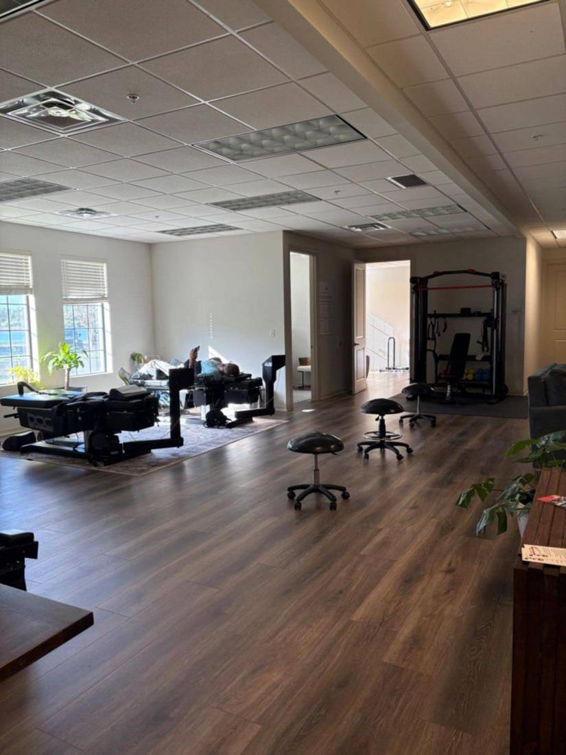
When a patient comes to Spine and Wellness Center Lakewood Ranch with this type of presentation, the exam protocol covers both systems:
Cervical spine assessment: Range of motion in all six directions, Spurling and distraction tests, dermatomal sensory testing, deep tendon reflex testing at C5-C7, and assessment of any upper extremity motor weakness that corresponds to a specific root level.
Shoulder assessment: Active and passive range of motion, rotator cuff strength testing (supraspinatus, infraspinatus, subscapularis), impingement provocation tests, and palpation of the AC joint and bicipital tendon groove.
Posture and thoracic outlet: Forward head posture and thoracic kyphosis directly load the cervical discs and foramina. We assess the upper thoracic region as part of any cervical presentation. Thoracic outlet maneuvers are included when arm symptoms extend past the elbow.
Digital X-ray is used when the history or exam suggests degenerative disc disease, prior trauma, or scoliosis that may be contributing. In our experience, a careful functional exam provides substantial diagnostic clarity before any imaging is needed, which matters both practically and in terms of unnecessary radiation exposure.
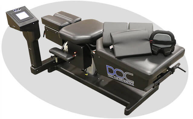
Once the exam identifies the structural drivers, care is targeted accordingly. Cervical-origin pain responds well to cervical adjustment, spinal decompression when a disc is implicated, and soft-tissue work to the surrounding musculature. Class IV laser therapy is used to reduce the inflammatory load around the compressed nerve root. Shoulder impingement, when confirmed, follows a separate protocol focused on reducing subacromial friction and restoring proper scapulothoracic mechanics.
For patients who have both problems simultaneously, care is layered and sequenced, addressing whichever component is most acutely limiting and progressing from there.
When to Get Evaluated
If you have been managing what you thought was a shoulder problem for more than 4 to 6 weeks without meaningful improvement, or if your shoulder pain is accompanied by neck stiffness, arm tingling, or numbness in specific fingers, those are strong reasons to have the cervical spine included in the assessment.
The two most common outcomes from a comprehensive exam are: (1) the neck is the primary driver and the shoulder has been incorrectly targeted, or (2) both systems are involved and treating only one will produce only partial relief. Either way, identifying the actual source changes the direction of care and the speed of recovery.
If you are in Lakewood Ranch, Bradenton, or Sarasota and you have this type of shoulder-neck presentation, call us at (727) 213-2982 or book directly at celluron.janeapp.com. The evaluation takes one visit and gives you a clear answer on where the pain is actually coming from.
For more on neck-related pain and headaches that trace to the cervical spine, see our neck pain and headaches page. For patients whose shoulder and neck symptoms developed after a car accident, our auto injury care page covers how these patterns are evaluated under Florida PIP.