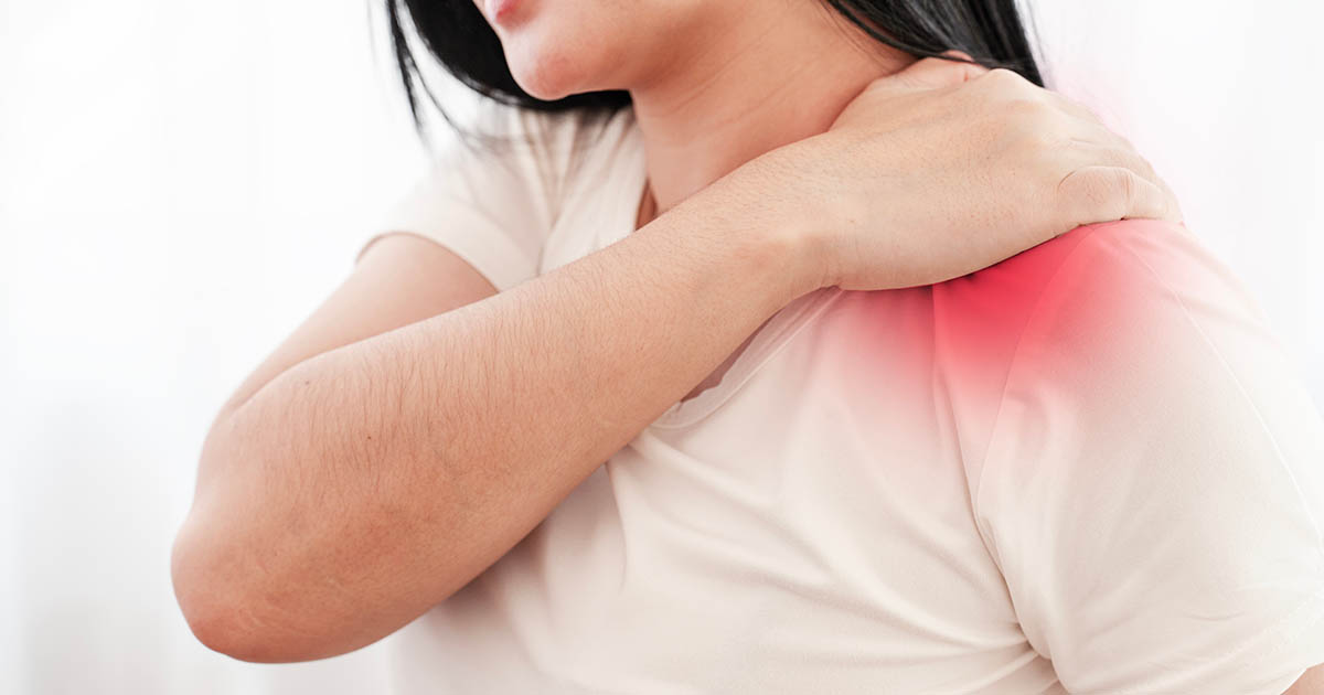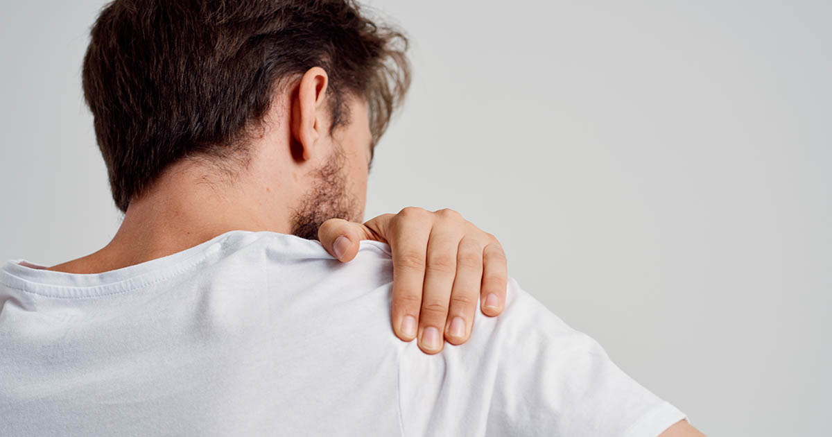
Shoulder pain is one of the most common complaints we evaluate at our Lakewood Ranch clinic, and the two diagnoses that come up most often are rotator cuff tendinitis and rotator cuff tear. Patients regularly come in having already diagnosed themselves online, convinced they either have "just inflammation" or "a tear that probably needs surgery." Most of the time, they are wrong in one direction or the other.
The clinical reality is more nuanced. Some tears are asymptomatic and require no intervention. Some cases of tendinitis that feel minor are actually the early presentation of a tear. And quite a few patients who have been told surgery is necessary actually have tendinopathy that responds well to conservative care. Getting the diagnosis right is the first step. This post walks through how to do that.
What the rotator cuff actually is
The rotator cuff is a group of four muscles (and their associated tendons) that surround the shoulder joint and hold the humeral head, the ball of the upper arm bone, seated against the glenoid, the shallow socket of the shoulder blade. The four muscles are:
- Supraspinatus (initiates arm elevation, most commonly injured)
- Infraspinatus (external rotation; often involved in overhead athlete injuries)
- Teres minor (assists infraspinatus in external rotation)
- Subscapularis (internal rotation; torn in falls on outstretched arm)
Together these four muscles allow the arm to rotate in all directions while keeping it in the socket. Without that dynamic stabilization, the joint would dislocate or impinge on surrounding structures with nearly every movement.
The tendons of these muscles converge to attach to the humeral head and pass through the subacromial space, a narrow corridor between the top of the humerus and the underside of the acromion (part of the shoulder blade). That corridor is where most of the trouble happens.
What is rotator cuff tendinitis?
Tendinitis (also written tendonitis; the terms are used interchangeably) means inflammation or irritation of the tendon. The structural integrity of the tendon is intact: the fibers are not torn. What you have is reactive tissue, often with some microdamage at the cellular level, that is inflamed and painful.
Rotator cuff tendinitis typically develops from one of three patterns:
- Repetitive overhead activity (swimming, tennis, painting ceilings, shelving) that loads the supraspinatus through its impingement zone over and over.
- Acute overload (a single heavy lift or awkward reach that irritates a previously normal tendon).
- Age-related degeneration (tendons lose vascularity and resilience with age; even ordinary activity can trigger tendinitis in a degenerated tendon).
Chronic tendinitis, where symptoms persist beyond 3 months, often transitions into tendinopathy: a degenerative state where the tendon has laid down disorganized collagen and is no longer truly inflamed but is structurally compromised. This distinction matters because anti-inflammatory approaches (ice, NSAIDs, cortisone) that help acute tendinitis can actually delay healing in chronic tendinopathy.
What is a rotator cuff tear?
A rotator cuff tear means the tendon fibers have actually separated. There are two broad categories:
Partial-thickness tears involve damage to some portion of the tendon but not all the way through. The tendon still has continuity. Many partial tears are found incidentally on MRI in people who have no significant symptoms. Others cause persistent pain that doesn't respond to conservative care.
Full-thickness tears (also called complete tears) go all the way through the tendon, disconnecting the muscle from its attachment. Small full-thickness tears often do not cause dramatic functional loss, especially in the early weeks. Large or massive tears, where multiple tendons are involved, typically produce significant weakness and limited range of motion.
How do tears happen? Two main mechanisms:
- Traumatic: A fall on an outstretched arm, a sudden jerk while lifting, or a shoulder dislocation. These can tear a previously healthy tendon. The pain is typically immediate and severe.
- Degenerative: A tear that develops gradually from accumulated microdamage in an already-compromised tendon. These often present with a history of shoulder pain that was "always kind of there" and then got noticeably worse.
The supraspinatus tendon tears more often than the other three combined. Its position in the subacromial space, where it is compressed with overhead movement, and its relatively poor blood supply at the critical zone near the attachment make it particularly vulnerable to both trauma and degeneration.
How to tell them apart without imaging
A good clinical history and physical exam can often distinguish tendinitis from a significant tear before imaging is ordered. Here is what matters.
Onset and history
Tendinitis typically develops gradually, often linked to a new activity or increased volume of an existing one. The pain may wax and wane with activity levels. A traumatic tear usually has an identifiable incident: a fall, a forceful lift, a sudden jerk. Degenerative tears often have no clear single event but a longer history of progressively worsening pain.
Strength testing
This is the most clinically useful separator. A significant full-thickness tear produces measurable weakness. The classic clinical test is the drop arm test: the examiner raises the patient's arm to 90 degrees abduction and asks them to slowly lower it. In a full-thickness supraspinatus tear, the arm drops rather than lowering smoothly. Pain alone does not mean there is a tear; weakness with specific testing is a much stronger indicator.
The empty can test specifically loads the supraspinatus: arm at 90 degrees forward flexion, thumbs pointed down (as if emptying a can). Resistance is applied downward. Pain or weakness in this position suggests supraspinatus involvement. Pain without weakness is more consistent with tendinitis; pain with weakness raises concern for a tear.
The painful arc
Both conditions often produce a painful arc: a range of motion, typically between 60 and 120 degrees of shoulder elevation, where pain is sharpest, corresponding to when the supraspinatus passes through the subacromial space. Tendinitis typically produces pain through the arc but maintains movement. A large tear often reduces the available arc on the affected side compared to the other shoulder.
Night pain pattern
Significant rotator cuff tears often produce marked nighttime pain that disrupts sleep, especially when lying on the affected shoulder. Tendinitis can also cause nighttime pain, but large tears tend to produce more severe sleep disruption because the tendon can no longer properly stabilize the shoulder even at rest.
This is also where distinguishing a rotator cuff problem from frozen shoulder (adhesive capsulitis) matters: frozen shoulder restricts passive range of motion (someone else moving your arm for you), while rotator cuff problems preserve passive motion. See that linked post for the full comparison.
When imaging changes the picture
Clinical testing is valuable, but definitive diagnosis requires imaging in most cases where a significant tear is suspected or conservative care is not working. Two modalities are used:
Ultrasound is dynamic, relatively inexpensive, and good at identifying full-thickness tears, fluid accumulation (bursitis), and calcific deposits. It can be performed in-office by experienced providers and allows the examiner to move the shoulder during imaging. Its limitations: it is highly operator-dependent and less reliable for partial-thickness tears.
MRI is the gold standard. It shows the full extent of any tear (size, retraction, muscle atrophy), the condition of the surrounding bursa, and the quality of the remaining tendon tissue. MRI is what guides the surgical decision when a tear is confirmed, because the size of the tear and the degree of muscle retraction and fatty infiltration determine whether surgical repair is feasible.
A point worth knowing: studies consistently show that partial-thickness tears and even some full-thickness tears are common incidental findings on MRI in people over 50 who have no shoulder symptoms. An MRI finding of "tear" is therefore a clinical diagnosis only when it matches the patient's symptoms and physical findings, not an automatic mandate for surgery.
Treating tendinitis without surgery
Rotator cuff tendinitis, especially when caught before it progresses to chronic tendinopathy, responds well to conservative care. The goal is to reduce the inflammatory load on the tendon while restoring the movement mechanics that allowed impingement to develop in the first place.
Shockwave therapy has emerged as one of the most effective non-surgical tools for tendinopathy, including calcific rotator cuff tendinitis (where calcium deposits develop in the tendon). High-intensity shockwave creates microtrauma at the tissue level that stimulates a healing response, can break down calcific deposits, and encourages angiogenesis, new blood vessel formation, in the notoriously hypovascular critical zone of the supraspinatus tendon. Many patients report improvement within 3 to 5 sessions.
Class IV laser therapy drives photons into the subacromial space through skin and muscle to reduce inflammation, promote mitochondrial activity in compromised tendon cells, and accelerate tissue repair. It works particularly well in the acute and subacute phases when genuine inflammation is present, and can be combined with shockwave for chronic tendinopathy.
Chiropractic evaluation and adjustment of the cervical spine and thoracic spine often plays a role that patients don't expect. Restricted mobility in the upper thoracic spine changes how the shoulder blade moves during arm elevation, a factor called scapular dyskinesis that loads the supraspinatus through an abnormal path. Restoring thoracic mobility and improving scapular mechanics takes pressure off the tendon even before direct shoulder work begins.
Specific rehabilitation exercises to strengthen the rotator cuff and periscapular muscles are always part of the long-term plan. The distinction between strengthening approaches for tendinitis versus a confirmed tear is important: loading a healthy tendon through its range of motion to stimulate collagen remodeling works well for tendinopathy, but eccentric and high-load programs need to be scaled carefully if a tear is present.
Non-surgical options for a confirmed rotator cuff tear
The question patients ask most often after hearing the word "tear" is: does this require surgery? For many tears, the honest answer is no, at least not initially. Research on rotator cuff tears, particularly partial-thickness tears and small full-thickness tears in patients over 60, consistently shows that structured conservative care produces functional outcomes comparable to early surgical repair in a meaningful proportion of patients.
The factors that push toward surgical evaluation:
- Acute traumatic full-thickness tears in younger patients (under 60) where the tendon is healthy and retraction is minimal
- Massive tears with significant retraction and early muscle atrophy (fatty infiltration of the muscle reduces the likelihood of successful surgical repair over time, so timing matters)
- Complete loss of active elevation (inability to raise the arm at all)
- Failure of at least 3 to 6 months of structured conservative care
For tears that do not meet those thresholds, the non-surgical pathway involves the same core tools as tendinitis care, with modifications:
Shockwave can be used to stimulate the tissue around a partial tear. Class IV laser supports the cellular environment of the remaining tendon fibers. Exercise loading is programmed carefully to avoid further tearing while building the compensatory strength needed from the intact portions of the cuff. The goal is functional restoration, not structural repair: the tear may not close, but the shoulder can often return to high function anyway.
Cortisone injections, while commonly offered, should be approached carefully with confirmed tears. Short-term pain relief is real, but cortisone has documented collagen-weakening effects on already-compromised tendon tissue, and repeated injections near an existing tear raise the concern of accelerating structural damage. We generally advise patients to use cortisone injections sparingly, and not as a substitute for addressing the underlying mechanics.
What to do next
The distinction between tendinitis and a rotator cuff tear matters for two reasons: it changes the treatment approach, and it changes what recovery realistically looks like. Treating a tear like uncomplicated tendinitis, particularly with aggressive loading early, can worsen it. Treating tendinopathy with excessive caution delays the specific stimulation that actually drives healing.
If your shoulder has been hurting for more than 4 to 6 weeks, or if you notice weakness with overhead movement or reaching behind your back, the right step is a clinical evaluation, not a wait-and-see approach. A proper exam, with orthopedic provocative testing and a clear functional assessment, can tell you in a single visit which category you are in and what the appropriate next step is.
At our Lakewood Ranch clinic, Dr. Banman performs a thorough shoulder evaluation including strength testing, range-of-motion measurement, and orthopedic provocation tests. If imaging is warranted based on the clinical findings, he will refer you for MRI or ultrasound and review the results with you directly. From there, a clear care plan is built around what your shoulder actually needs, not a generic protocol.
For more on how we treat shoulder-related conditions, see our frozen shoulder page and our overview of shockwave therapy for tendinopathy.