Most people associate knee pain with a specific event: a collision on the pickleball court, a bad landing on a run, a slip on wet pavement. But a large percentage of patients who come in with knee pain cannot point to any moment when it started. It was just there one morning, or it built up gradually over weeks, or it showed up after a long stretch of sitting at a desk. No trauma. No incident. Just pain.
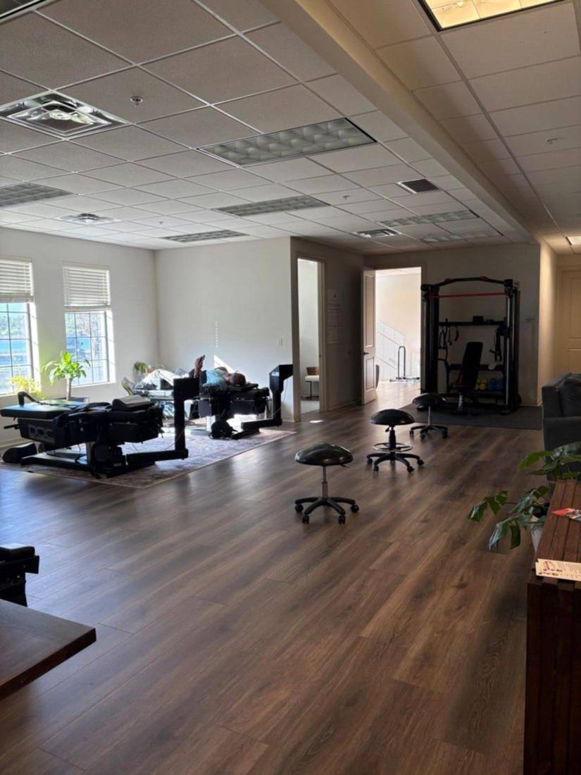
That presentation confuses people because the common mental model of injury requires a cause-and-effect story. Without the story, some patients assume they must have done something they didn't notice, or that the pain will resolve on its own. In our experience at Spine and Wellness Center Lakewood Ranch, that second assumption is the one that leads to months of unnecessary waiting.
Knee pain without a clear traumatic event is not a mystery condition. It has specific, identifiable causes. This article covers the most common ones, how to tell them apart by symptom pattern, and what kinds of care actually move the needle.
Why the Knee Is Vulnerable to Non-Traumatic Pain
The knee is structurally complex and mechanically demanding. It supports your full body weight through most of daily life, it bends and straightens hundreds of times a day, and it depends heavily on the surrounding soft tissue (muscles, tendons, bursae, the joint capsule) for stability because the bony structure itself offers relatively little. When any of those supporting structures is overloaded, irritated, or getting inadequate neurological input, pain develops without any single event to blame.
Several factors make the knee particularly susceptible to this kind of gradual-onset pain:
- Imbalanced hip and quad strength shifts load onto the joint rather than the muscles around it
- Prolonged sitting shortens the hip flexors and changes how the femur rotates during walking, which alters knee mechanics
- Lumbar spine dysfunction can refer pain directly down the leg to the knee region
- Peripheral nerve compression anywhere along the sciatic or femoral nerve pathway can produce burning, aching, or pressure sensations that are felt at the knee
- Progressive cartilage loss happens without symptoms for years, then crosses a threshold where weight-bearing becomes painful
The most useful question when evaluating knee pain without injury is not "what did you do to it" but "what does the pattern of the pain tell us about the structure involved." That distinction often points to the answer in a single exam visit.
Cause 1: Referred Pain from the Lumbar Spine or Hip
This is the cause most patients don't consider and most imaging studies miss. The lumbar spine and hip joint both share nerve pathways that refer pain into the thigh and knee region. When a lumbar disc at L3-L4 is compressed or a facet joint at that level is irritated, the pain can be felt primarily in the inner knee or lower thigh, with minimal or no lower back awareness at all. The patient feels a knee problem. The source is the spine.
Similarly, hip joint pathology, including early hip arthritis, labral issues, and piriformis syndrome, commonly refers pain down into the knee region. The hip and knee share femoral and obturator nerve branches, so when the hip is the primary problem, the knee often carries the symptom load while the hip itself remains relatively quiet.
The distinguishing feature of referred knee pain is that it tends not to be provoked by direct pressure on the knee structures themselves. Pressing on the medial joint line, the patellar tendon, or the lateral knee doesn't reproduce the familiar pain the way it does in a localized knee problem. Instead, reproducing the pain requires loading the spine (standing, walking, bending) or moving the hip through range of motion.
A thorough evaluation should always include lumbar and hip assessment when the knee is the presenting complaint. Treating the knee while the spine or hip is the actual source produces limited results.
For patients whose knee pain follows a pattern consistent with nerve root compression, our sciatica evaluation and lumbar spine assessment can identify whether the lumbar spine is contributing to what appears to be a knee problem.
Cause 2: Patellofemoral Syndrome
Patellofemoral syndrome (PFS) is one of the most common knee complaints in adults of all ages, and it frequently develops without any traumatic event. The patella (kneecap) needs to track smoothly in the trochlear groove of the femur as the knee bends and straightens. When the muscular balance around the knee and hip is off, the patella tracks improperly. This creates abnormal pressure on the underside of the kneecap and produces pain, particularly with:
- Going up or down stairs
- Sitting for extended periods with the knee bent (the classic "theater sign")
- Squatting or kneeling
- Running or walking for sustained distances
The pain is typically diffuse and aching, often described as being "under" or "around" the kneecap. It is not usually sharp or point-specific. Most patients cannot identify an exact moment when it started. It came on gradually as activity demands changed, as sitting patterns shifted (common after transitions to desk work or travel), or as hip and quad strength declined with age or inactivity.
What drives patellofemoral syndrome is almost never the knee itself. The typical culprits are weak hip abductors and external rotators (especially gluteus medius) that allow the femur to internally rotate and adduct under load, combined with tight iliotibial band structures that pull the patella laterally. Addressing those proximal drivers produces better outcomes than focusing exclusively on the knee.
Cause 3: Iliotibial Band Syndrome and Bursitis
The iliotibial (IT) band is a dense strip of connective tissue that runs from the hip down the lateral thigh and attaches just below the lateral knee. When it becomes chronically tight or irritated, it creates friction over the lateral femoral condyle, producing a sharp or burning pain on the outside of the knee. This is IT band syndrome, and it is extremely common in people who walk frequently, do yard work, climb stairs regularly, or sit for long stretches with the hip and knee at 90 degrees.
No single injury event causes IT band syndrome. It develops from accumulated mechanical load combined with hip abductor weakness and inadequate soft tissue mobility.
Bursitis is a different mechanism but similarly non-traumatic in many cases. The knee has several bursae (fluid-filled sacs that reduce friction between structures). The prepatellar bursa in front of the kneecap and the pes anserine bursa on the inner knee below the joint line are two common sites. They can become inflamed through repetitive low-level friction, prolonged kneeling (common in gardening, flooring work, or prayer), or metabolic factors like gout and diabetes, neither of which requires an impact or twist to produce knee pain.
Pes anserine bursitis in particular is frequently encountered in patients with osteoarthritis and in those who are carrying extra weight, where the increased medial compartment load creates chronic bursal irritation without any identifiable injury.
Cause 4: Osteoarthritis Crossing a Threshold
Knee osteoarthritis (OA) is a progressive loss of cartilage, the smooth tissue that covers the ends of the bones inside the joint. Cartilage loss is painless for years because cartilage itself has no nerve supply. Pain emerges when the loss reaches a degree that changes the mechanics of the joint: the joint space narrows, bone begins to carry load it wasn't designed to bear directly, and the synovial membrane (the joint lining) becomes chronically inflamed.
This progression means many patients with meaningful knee OA have no history of knee trauma at all. The damage accumulated quietly over time. Then, often in the late 40s or early 50s, the knee begins to ache with weight-bearing, to stiffen after rest, to swell after activity, or to feel unreliable on uneven ground. There was no incident because the incident was twenty years of accumulated loading.
The key distinguishing features of OA-driven knee pain include:
- Morning stiffness that loosens up within 30 minutes of movement (unlike inflammatory arthritis, which stays stiff longer)
- Pain that worsens with weight-bearing activity and improves with rest (unlike patellofemoral pain, which is worse with prolonged sitting)
- Bony enlargement at the joint margins that can sometimes be felt on palpation
- Crepitus (grinding or crunching sensation) during movement
- Gradual onset over months or years, not weeks
Imaging confirms OA but does not predict symptoms well. Many patients with moderate OA on X-ray have manageable pain; others with mild radiographic changes are significantly limited. The functional picture matters more than the image alone.
Non-surgical options for knee OA have improved considerably. Approaches like shockwave therapy target the soft tissue component of knee pain and can reduce inflammation in the periarticular structures, improving function without surgical intervention.
Cause 5: Peripheral Neuropathy
Peripheral neuropathy is damage or dysfunction of the peripheral nerves, those outside the brain and spinal cord. When the nerves that supply the knee and lower leg are affected, the symptoms can present as burning, aching, pressure, or even a vague "deep bone pain" at the knee level. Patients sometimes describe it as a toothache-like sensation inside the joint, rather than the mechanical pain of a structural problem.
Neuropathic knee pain is frequently mistaken for joint pathology and sent down an orthopedic evaluation pathway that produces normal or near-normal imaging findings. The patient is told the knee "looks fine," which is accurate, because the problem isn't in the joint structures at all. The problem is in the nerve supply.
Common drivers of peripheral neuropathy that manifest at the knee include:
- Diabetes (the most common systemic cause of peripheral neuropathy)
- Lumbar disc compression of the L3-L4 nerve roots, which supply the anterior knee region
- Common peroneal nerve entrapment at the fibular head (lateral knee region)
- Saphenous nerve entrapment in the adductor canal (medial knee region)
- Systemic metabolic conditions including hypothyroidism and vitamin B12 deficiency
The pattern of neuropathic pain tends to be diffuse, sometimes bilateral, often worse at night or with sustained positions rather than with specific mechanical loading. It may be accompanied by tingling, numbness, or temperature changes in the foot or lower leg.
Our neuropathy evaluation uses both clinical testing and nerve function assessment to identify whether peripheral nerve dysfunction is driving the knee pain picture, rather than structural joint pathology.
Red Flags That Need a Prompt Medical Evaluation
Most non-traumatic knee pain is mechanical in origin and responds to conservative care. However, certain presentations warrant prompt evaluation beyond a chiropractic or musculoskeletal workup:
- Knee pain with significant, rapid swelling and warmth suggesting infection or inflammatory arthritis (rheumatoid, gout, pseudogout)
- Unexplained weight loss accompanying knee pain (could indicate a systemic or malignant process)
- Nighttime pain that is constant and not related to position (classical red flag for non-mechanical causes)
- Knee pain in a person with a history of cancer, particularly if the pain is persistent and unresponsive to any intervention
- Significant locking of the knee (inability to fully extend), which can indicate a loose body or meniscal bucket-handle tear even without a clear injury event
If any of these features are present, a referral for imaging and specialist evaluation is the appropriate next step. Most patients presenting with non-traumatic knee pain do not have these features, but the distinction is important.
What a Good Evaluation Covers
A thorough evaluation for non-traumatic knee pain should not start and end with the knee itself. In our experience, the assessment needs to work through several layers:
- History: Pattern of symptoms (onset, timing, aggravating and relieving factors), bilateral vs. unilateral, presence of back pain, systemic conditions, activity history, sitting and postural habits
- Lumbar and hip screen: Range of motion, nerve tension tests (straight leg raise, femoral nerve tension), hip strength and mobility assessment
- Knee-specific exam: Joint line palpation, patellar mobility and tracking, ligament stress tests, bursal tenderness assessment
- Neurological screen: Sensation, reflexes, and if indicated, nerve function testing to rule out neuropathic contribution
- Functional movement: Single-leg squat, step-down test, gait observation to see how the knee is actually loading
Imaging (X-ray for bony structure, MRI for soft tissue) is a useful supplement when indicated, but many of the causes discussed here are diagnosed clinically and do not require imaging to begin treatment. Starting care while awaiting imaging is often appropriate when the presentation points clearly to a mechanical cause.
Non-Surgical Options Worth Knowing About
Many patients with unexplained knee pain have spent time on NSAIDs, tried rest, and possibly had a cortisone injection that helped temporarily. These approaches address symptoms rather than the underlying mechanical or neurological drivers.
Non-surgical options with better long-term rationale for the causes described here include:
- Chiropractic care for lumbar and hip mechanics: When the spine or hip is the source of referred knee pain, addressing joint mobility and nerve root irritation at that level is the logical starting point. Treating the knee while the spine remains the driver produces incomplete results.
- Shockwave or softwave therapy: Useful for the tendon and bursal components of knee pain, including IT band syndrome, patellar tendinopathy, and pes anserine bursitis. These therapies stimulate tissue repair and reduce localized inflammation in soft tissue structures.
- Targeted rehabilitation: Hip abductor and external rotator strengthening to correct the proximal mechanics that drive patellofemoral syndrome. This is not generic "leg exercises"; it requires identifying which deficits are present and addressing them specifically.
- Neuropathy protocols: For patients whose knee pain has a significant neurological component, nerve-directed therapies can reduce the sensory disturbance and improve function in ways that standard musculoskeletal care does not.
For patients curious about whether their knee pain has a referred or spinal component, our overview of hip and sciatic-pattern pain covers how referred lower extremity pain is evaluated and what distinguishes it from local joint pathology.
A Note on Waiting It Out
One of the most common patterns we see is a patient who waited six months, twelve months, or longer before seeking evaluation, assuming the knee would "sort itself out." For some presentations, particularly mild patellofemoral irritation from a temporary activity change, that is reasonable. For most of the causes discussed in this article, waiting tends to allow the compensatory patterns to worsen rather than resolve.
When the lumbar spine is referring pain to the knee, that disc or facet is not improving during the waiting period. When IT band syndrome or bursitis is the driver, the accumulated mechanical irritation doesn't self-correct without addressing the muscle imbalances behind it. When osteoarthritis is the cause, the window for conservative management is not unlimited: early-to-moderate OA responds much better to non-surgical care than advanced OA does.
The earlier the structural source is identified, the more options exist for managing it without escalating to injections or surgical consultation.
Summary
Non-traumatic knee pain has identifiable causes. The five most common are referred pain from the lumbar spine or hip, patellofemoral syndrome driven by hip and quad mechanics, IT band syndrome and bursitis from accumulated soft tissue load, osteoarthritis crossing a symptomatic threshold, and peripheral neuropathy affecting the nerve supply to the knee region. Each has a distinct symptom pattern and a distinct treatment rationale.
The evaluation approach matters. Starting with only the knee, without examining the spine, hip, and neurological system, misses the cases where the knee is the recipient of the problem rather than the source. In our experience at Spine and Wellness Center Lakewood Ranch, getting the anatomy right changes everything about what care is actually warranted.