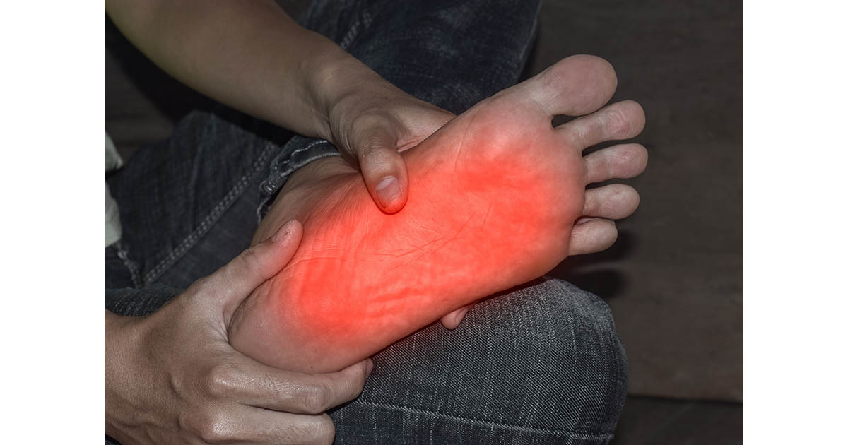

Foot drop is the inability to lift the front part of your foot when you step. The foot hangs down or drags, making a normal walking stride impossible. Most people dismiss the first few episodes as fatigue or a bad shoe. By the time they recognize the pattern, weeks have passed, and the underlying nerve compression has had time to deepen.
At our Lakewood Ranch clinic, foot drop is one of the presentations we take seriously from the very first visit. In more than 23 years of practice, the pattern is consistent: the majority of cases we see start with a structural problem in the lower lumbar spine, specifically compression of the L4 or L5 nerve root. The good news is that when the cause is spinal, early conservative care can make a real difference. Understanding why your foot drops is the first step toward getting that care. If you have been dealing with nerve-related symptoms in your legs, our neuropathy condition page covers the broader picture of how nerve damage presents and progresses.
What Foot Drop Actually Is
The medical term is "drop foot" or "foot drop," and it describes weakness or paralysis of the muscles that lift the foot at the ankle. That movement is called dorsiflexion. When the tibialis anterior and the peroneal muscles cannot fire properly, the foot cannot clear the ground during the swing phase of walking.
The compensation gait that develops is called a steppage gait: you hike your hip and knee high to swing the affected leg forward, almost like climbing a step that is not there. It is tiring, and it dramatically increases fall risk. On stairs, curbs, or uneven ground, a dragging foot catches easily.
Foot drop is a symptom, not a diagnosis. The question that matters is: what nerve is not getting its signal through, and why?
The Most Common Cause: L4-L5 Disc Herniation
The nerve that controls dorsiflexion runs from the L4 and L5 levels of your lumbar spine, through the pelvis, down the outside of the knee (as the common peroneal nerve), and into the foot. A herniated or bulging disc at L4-L5 presses directly on the L5 nerve root. That compression disrupts the signal before it ever reaches the peroneal nerve downstream.
This is why foot drop and herniated disc so frequently appear together. A patient comes in reporting that their "sciatica" has been bad lately and they have been tripping more. On exam, testing ankle dorsiflexion strength tells us immediately that L5 is being compromised, not just irritated. The distinction matters for how we plan care.
L4 root compression can produce a similar picture, since L4 also contributes to tibialis anterior function. Both levels are in the disc-heavy zone of the lower back, the area responsible for most structural back pain in adults over 40.
In our practice, foot drop that develops alongside low back pain or buttock pain almost always has a spinal component. We look for L4-L5 involvement first because that combination is far more common than isolated peroneal nerve damage at the knee.
Other Causes Worth Knowing
A spinal disc is the most common culprit, but not the only one. Other causes fall into three categories:
Spinal stenosis at L4-L5
When the spinal canal narrows (stenosis), it can compress the nerve roots just as a disc can. The difference is that spinal stenosis tends to produce symptoms that worsen with standing and walking and improve with sitting or bending forward. If your foot drop or leg weakness gets notably worse after walking a block and eases when you sit on a bench, stenosis is worth investigating specifically.
Peroneal nerve compression at the knee
The common peroneal nerve wraps around the outside of the knee just below the joint. It can be compressed by: crossing your legs habitually for long periods, wearing a tight knee brace, spending extended time kneeling or squatting, or a direct blow. This peripheral compression produces foot drop without any back pain, which sometimes leads patients to assume the spine is not involved, and in this case they are right. Sorting out where along the nerve the problem sits is exactly what a clinical exam is designed to do.
Peripheral neuropathy
Advanced peripheral neuropathy, from diabetes, B12 deficiency, or other metabolic causes, can damage the peroneal nerve fibers as part of a broader nerve failure pattern. In these cases, foot drop is usually accompanied by other neuropathy signs: burning, tingling, or numbness in both feet, often symmetric. If you recognize that picture, our neuropathy program addresses the multi-system approach that metabolic nerve damage requires.
Central nervous system causes
Stroke, multiple sclerosis, and brain or spinal cord tumors can cause foot drop by disrupting the upper motor neuron pathways. These causes are less common but critical to rule out, especially if the foot drop came on suddenly, if there is facial weakness or speech changes, or if both sides are affected. Any sudden-onset foot drop without a clear mechanical cause warrants urgent neurological evaluation, not a wait-and-see approach.
When Foot Drop Is an Emergency
Two presentations require same-day emergency evaluation. If either applies to you, go to an emergency room or call 911, not a chiropractic office:
- Sudden onset with bladder or bowel changes. If you lose control of your bladder or bowel at the same time foot drop develops, this may be cauda equina syndrome, a compression of the nerve bundle at the base of the spinal cord. It is a surgical emergency.
- Sudden onset with other neurological signs. New foot drop plus facial drooping, speech difficulty, arm weakness, or vision changes points toward a central event (stroke or similar). Do not drive yourself. Call emergency services.
When foot drop comes on gradually, over days to weeks, and is associated with back pain or a known disc problem, the urgency level is lower but still real. Progressive neurological deficits that are left untreated can become permanent.
How We Evaluate Foot Drop at Our Clinic
A thorough clinical exam tells us more than most patients expect, even before any imaging. We test:
- Ankle dorsiflexion strength (ask you to pull your toes up against resistance)
- Big toe extension strength (L5 specific)
- Deep tendon reflexes at the knee and ankle (help differentiate L4 from L5 from peripheral)
- Sensation mapping along the L4 and L5 dermatomes (the outside of the lower leg and the top of the foot)
- Straight leg raise to assess nerve root tension
- Gait observation (the steppage pattern is often visible immediately)
From those findings, we can usually identify whether the lesion is at the spine, at the knee, or is part of a diffuse peripheral pattern. If imaging is indicated, we refer for MRI of the lumbar spine or nerve conduction studies. We coordinate with your neurologist or orthopedic spine specialist when the findings suggest surgery is worth evaluating. Conservative care and surgical evaluation are not mutually exclusive, and knowing which path fits your case is something we can help you figure out.
What Conservative Treatment Looks Like
When the cause is a herniated disc or spinal stenosis at L4-L5, the goal of conservative care is to reduce the pressure on the nerve root and give the nerve the environment it needs to recover its signal. Our approach involves several tools, combined based on your specific presentation:
Spinal decompression
Non-surgical spinal decompression is the treatment we reach for most often in disc-driven foot drop. The decompression table creates controlled traction at the L4-L5 level, reducing intradiscal pressure and allowing bulging material to migrate away from the nerve root. Many patients in this situation report changes in their foot-lifting ability within several weeks of consistent treatment, though individual responses vary considerably.
Chiropractic adjustments
Targeted adjustments at the lumbar spine address joint restrictions that compound nerve root irritation. A fixated facet joint at L4-L5 does not cause disc herniation on its own, but it can worsen the mechanical environment around an already-compressed nerve. Restoring segmental motion takes some of that compounding stress off the root.
Class IV laser therapy
Laser therapy at the therapeutic level we use promotes circulation to compressed nerve tissue and reduces local inflammation. For peripheral neuropathy cases contributing to foot drop, laser along the peroneal nerve pathway is part of our nerve-recovery protocol.
Electrical muscle stimulation
When the tibialis anterior is genuinely weakened from nerve compromise, EMS can maintain some muscle activation while the nerve recovers. It does not replace the nerve signal, but it helps prevent the atrophy that makes recovery harder once the nerve is freed. Combined with exercises targeting ankle dorsiflexion, this approach keeps the affected muscles engaged.
Ankle-foot orthosis (AFO) referral
For patients with significant foot drop, we coordinate a referral for an AFO, a light brace that holds the foot at 90 degrees during walking. The brace does not treat the cause, but it prevents falls and keeps the patient mobile while the spinal or nerve treatment has time to work. Safety during recovery is part of the plan.
Recovery: What to Expect
Foot drop recovery depends heavily on how long the nerve has been compressed and how much signal is still getting through. A nerve root that has been squeezed for two weeks behaves differently than one that has been compressed for two years. Early intervention, before the nerve loses its protective myelin sheath, gives the best window for recovery.
In our experience, patients with disc-driven foot drop who start decompression-based care within the first few months of onset often report gradual improvement in dorsiflexion strength over 8 to 16 weeks, though individual results vary and some cases require longer timelines or surgical intervention when the compression is severe. Nerve tissue heals slowly, roughly 1 mm per day along the fiber, so patience is not optional. What matters is that the pressure is off and the nerve has the environment it needs to repair itself.
Foot drop from peripheral neuropathy tends to respond more slowly and is more dependent on managing the underlying metabolic cause. When the root problem is diabetes, for instance, blood sugar control is as important as any local treatment. We look at the full picture, not just the foot.
If you are in the Lakewood Ranch or Bradenton area and have noticed changes in how you walk, tripping more often, or difficulty lifting your foot, do not wait until the symptom becomes a fall. An evaluation takes one appointment and gives you a real answer about what is driving it and whether conservative care applies to your case.