
The report lands in your patient portal at 11pm. You type the phrase into Google. Fifteen minutes later you are reading about nerve damage and surgical fusions. That spiral is almost always premature.
"Loss of cervical lordosis" is one of the most common findings on neck X-rays and MRIs in Lakewood Ranch patients we see for chronic neck pain and headaches. It shows up in people who have been staring at laptops for years, people who had a rear-end collision a decade ago, and occasionally in teenagers who've spent most of high school looking down at a phone. It is not a sentence. It is a starting point.
Here is what the finding actually means and what the evidence says you can do about it without a scalpel.
What cervical lordosis actually is
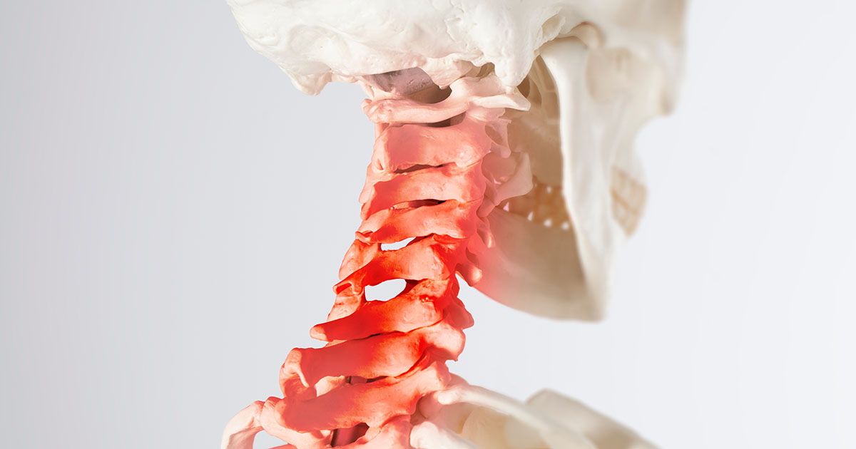
Your cervical spine (the seven vertebrae that make up your neck, C1 through C7) is supposed to have a gentle backward "C" curve when viewed from the side. Radiologists measure this curve by the Cobb angle method: they draw lines along the endplates of C2 and C7 and measure the angle where those lines meet.
A healthy cervical lordosis typically measures somewhere between 20 and 40 degrees of curvature. That curve is not cosmetic. It functions as a shock absorber for the approximately 10 to 12 pounds your head puts on your neck every time you hold it upright. When the curve is intact, that load is distributed across the discs and facet joints in a balanced way. When the curve flattens, the load shifts forward and concentrates on the front edges of the vertebrae and the discs between them.
Over time, that concentrated load can accelerate disc compression and facet degeneration. A neck with less than 20 degrees of curvature is generally described as having "loss of lordosis." A neck that has no curve at all is called "straight neck" or "military neck" (the latter because the rigid upright posture resembles the stance of a soldier at attention). A neck that has actually reversed into a forward curve is called "kyphotic," and that is the stage that warrants the most attention.
Loss of cervical lordosis is a structural finding, not a diagnosis on its own. Many people have a straightened cervical curve and no symptoms. Many people with significant symptoms have a curve that looks acceptable on imaging. Imaging tells us what the structure looks like; it does not tell us why you hurt or what to do about it. That part requires an examination.
What the imaging report means in plain language
When a radiologist writes "loss of cervical lordosis" or "straightening of the normal cervical lordosis," they are describing a curve that has decreased but has not reversed. "Reversal of cervical lordosis" or "cervical kyphosis" means the curve has flipped forward. These are different findings with different implications.
Sometimes you will see phrases like "this may represent muscle spasm" in the report. That is radiologist-speak for: we cannot tell from a static image whether this is a structural change that has accumulated over years, or a temporary splinting response your neck muscles create when they are in pain or guarding. After a car accident, for example, the paravertebral muscles tighten reflexively and can pull the cervical spine into a straight position even if the underlying curve is fine. The curve may normalize once the acute muscle spasm resolves.
What the report typically cannot tell you: how long this has been present, what caused it, whether it is progressing, or whether it is the primary driver of your symptoms. That is why the imaging report is an input to the examination, not a replacement for it.
How the cervical curve gets lost
The two most common causes we see in our Lakewood Ranch office are sustained forward head posture and prior whiplash-mechanism injuries. They often overlap.
Forward head posture (FHP). For every inch your head drifts forward of your shoulders, the effective weight on your lower cervical discs and muscles increases by roughly 10 pounds. Someone sitting with their head 3 inches forward of center is putting an estimated 42 pounds of load on a structure designed for 12. Over months and years, the muscles on the back of the neck (the suboccipital group, the upper trapezius, the levator scapulae) shorten and tighten. The muscles on the front of the neck weaken. The discs at C5-C6 and C6-C7 bear the brunt. Tech neck is the accelerant of this process for most working adults.
Whiplash-mechanism injury. Even a low-speed rear-end collision puts rapid extension-then-flexion forces on the cervical spine. The paravertebral muscles can go into protective spasm and, if the injury is not properly treated, stay there for months. Scar tissue forms in the soft tissues. The motion segments at C4-C5 through C6-C7 begin to stiffen. Years later, the imaging shows a flattened curve and the patient cannot recall any "serious" injury because the collision happened at parking-lot speed and "not a big deal" was the impression at the time.
Other contributors include:
- Degenerative disc disease at multiple cervical levels (the loss of disc height changes segment angles)
- Osteoporosis-related vertebral changes in older women
- Sleeping positions that force the neck into sustained flexion (face-down with the head rotated hard to one side)
- Occupations that require prolonged looking downward: surgeons, dentists, jewelers, seamstresses
What symptoms does it cause, and what does it not cause
Loss of cervical lordosis is associated with neck pain, upper trapezius tension, headaches that start at the base of the skull, and reduced range of motion. It can also contribute to earlier facet joint wear and disc degeneration at the flattened segments, which over time may produce symptoms from nerve root irritation or compression.
However, it does not directly cause arm pain, hand numbness, or weakness on its own. Those symptoms point toward nerve root involvement or, in more serious cases, spinal cord involvement. If you have a flattened cervical curve AND arm symptoms that radiate past your elbow AND grip weakness AND balance problems, that combination warrants urgent evaluation because it can indicate early cervical myelopathy. See a provider for same-week evaluation if that is your picture.
For the much more common pattern (flattened curve plus neck stiffness, midline neck pain, and occipital headaches with no arm symptoms), the prognosis is generally favorable with appropriate conservative care.
What chiropractic care and decompression can actually do
The goal is not to "fix the curve" in one visit, or even in a month. The cervical lordosis developed over years. Structural changes take time, consistency, and the right mechanical inputs. Here is what the evidence supports for the non-surgical approach:
Chiropractic adjustments to the cervical and upper thoracic spine. Segmental manipulation improves joint mobility at the stiff levels, reduces facet capsule irritation, and resets the muscle tone patterns that are reinforcing the flattened posture. At our Lakewood Ranch office, Dr. Banman has over 23 years of clinical experience identifying which specific segments need to move and in which direction. Not every approach is the same: a C4-C5 manipulation is different from a C6-C7 manipulation, and the thrust vectors matter.
Cervical traction and non-surgical spinal decompression. Specific traction angles can apply an unloading force that gently encourages extension at the flattened segments. A study published in the European Spine Journal found that sustained mechanical traction combined with a structured exercise program produced measurable improvement in cervical lordosis angles compared to exercise alone. The key word is "structured" - random traction with no guidance on angle or position tends to do little.
Posture correction and deep cervical flexor training. The longus colli and longus capitis (the muscles running along the front of the cervical vertebrae) are almost always inhibited and weakened in patients with lost cervical lordosis. Reactivating them with specific low-load endurance exercises is one of the most evidence-supported interventions for both pain and curve restoration. These are not the exercises most people do at the gym. They require instruction and feedback to do correctly.
Class IV laser therapy. For patients who have accompanying disc-related inflammation at the flattened segments, photobiomodulation (Class IV laser) can reduce inflammatory cytokines in the disc and surrounding soft tissue. Many patients in our practice report significant reduction in stiffness and pain responsiveness after a course of laser combined with adjustments, which makes the structural work more productive.
What a realistic recovery trajectory looks like
For a patient with mild to moderate loss of cervical lordosis, no arm symptoms, and pain primarily in the neck and occipital region, our experience with this population over 23 years is this: most people feel meaningfully better within 4 to 6 weeks of consistent care. Range of motion typically improves faster than pain resolves. The structural change in the curve takes longer; a meaningful restoration of curve angle (10 to 15 degrees on follow-up X-ray) generally requires a minimum of 3 to 6 months of consistent care with home exercises.
Some patients achieve good pain relief and functional improvement without complete curve restoration. Others restore the curve gradually over a year of care and maintenance. The two outcomes are not mutually exclusive.
What does not produce results: one or two adjustments followed by a long gap, no home exercise program, continuing the same posture patterns that created the problem, and sleeping face-down. If you are not addressing the daily mechanical input, structural correction is an uphill fight.
What we look for at the initial exam: range of motion in all six planes, orthopedic and neurological testing to rule out nerve root involvement, and a lateral cervical X-ray with measurement if one has not been taken recently. From that picture, Dr. Banman can tell you specifically which levels are restricted, whether the curve loss is segmental or global, and what a realistic care plan looks like for your situation.
If you have been sitting on a confusing imaging report or have had neck pain and headaches that keep coming back despite stretching and over-the-counter remedies, a structured evaluation is the fastest way to get a clear answer. We see new patients at our Lakewood Ranch office within 24 to 48 hours of calling, and the initial visit includes a thorough review of any existing imaging you bring in. If you have been evaluated for cervical radiculopathy and the arm symptoms have resolved but the neck stiffness remains, loss of lordosis is often what is left driving the picture.