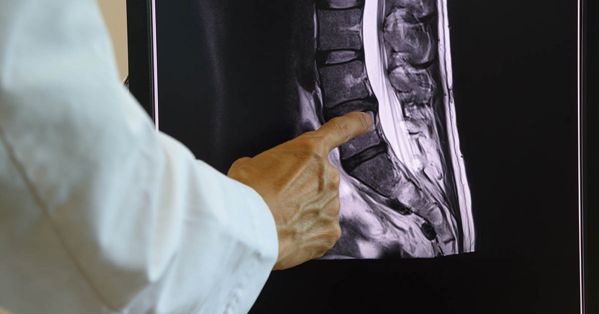
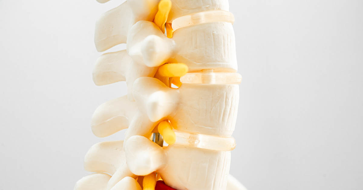
Every week someone comes into our Lakewood Ranch clinic asking the same question: "Should I get an X-ray first?" They have already looked it up online, read conflicting advice, and are wondering whether skipping imaging means missing something serious. The honest answer is that the question itself reflects how medicine has changed. Guidelines from the American College of Physicians and the North American Spine Society both say the same thing: for most acute lower back pain, imaging in the first six weeks rarely changes the initial care plan. What matters more than an X-ray is a thorough physical exam.
That said, there are specific situations where lumbar X-rays or advanced imaging genuinely moves the needle. Knowing the difference is what separates an informed patient from one who either delays necessary imaging or who goes through unnecessary radiation exposure for something that was going to resolve on its own.
Why chiropractors do not order X-rays on every back pain patient
This surprises some patients. Twenty-three years of clinical practice will teach you that lumbar spine films often show changes that look alarming on paper but correlate poorly with pain. A 2015 study in the European Journal of Radiology found degenerative disc changes on MRI in 52 percent of asymptomatic 30-year-olds and in more than 80 percent of asymptomatic 55-year-olds. People walking around without any back pain at all had findings that sound dramatic on a radiology report.
Ordering an X-ray on everyone who walks in with back pain does not just expose them to unnecessary radiation. It generates findings that can scare a patient, lead to over-treatment, and sometimes lead to surgery for something that was not actually causing the pain. The clinical exam tells us far more about what is actually going on than a picture that shows anatomy without telling us which structure hurts and why.
At the same time, a targeted history and physical exam can flag cases where imaging is not optional. Those cases get films the same week, sometimes the same day.
The red flags that move imaging to the top of the list
In chiropractic and primary care alike, certain findings during the history and exam escalate imaging from "not urgent" to "this week" or "now." None of these require a patient to diagnose themselves; they are things the clinician is screening for. But understanding them helps explain why two patients with similar pain levels might get very different recommendations.
- Age over 50 with new-onset back pain. Degenerative changes accelerate after 50, and the risk of compression fractures from osteoporosis rises significantly, especially in women who have not been screened for bone density loss. New pain in this age group without a clear mechanical trigger is worth imaging before we start any loading or manipulation.
- History of cancer. Spinal metastases can present as back pain before anything else is obvious. Any patient with a history of cancer who develops back pain gets imaging, full stop. We refer out when indicated.
- Unexplained weight loss or fever alongside back pain. These two together are the classic presentation for spinal infection (discitis or vertebral osteomyelitis) or, less commonly, a paraspinal abscess. Pain from these conditions does not respond to chiropractic care and can deteriorate rapidly.
- Recent significant trauma. A fall from height, a motor vehicle crash, or a direct blow to the spine in an older adult or anyone on blood thinners warrants imaging to rule out fracture before any hands-on care.
- Bowel or bladder dysfunction alongside back pain. This combination is a neurological emergency, potentially cauda equina syndrome, and goes to the emergency room before it goes anywhere else. I tell every patient this on intake. If you ever lose control of your bladder or bowels along with back pain, that visit to our office waits.
- Severe or progressive neurological deficit. A foot drop that develops over hours, or rapidly worsening leg weakness that accompanies back pain, is a different clinical picture than the usual sciatica presentation. It needs imaging before conservative care begins.
- Pain that is completely unresponsive to positional changes and worse at night. Disc pain and facet pain both shift with movement and position. Pain that is constant, unrelenting, and actually worse when you lie down can signal a non-mechanical cause (inflammatory arthropathy, tumor, or infection) that imaging will clarify.
- Long-term steroid use or osteoporosis diagnosis. Chronic steroid use causes accelerated bone density loss. Patients on prednisone or similar medications for more than three months are at meaningful risk for vertebral compression fractures even from minor loading events.
What a standard lumbar X-ray actually shows
When we do order films, a standard lumbar spine series typically includes anteroposterior (AP) and lateral views, sometimes with flexion-extension laterals if we are evaluating for instability. What we are looking at, specifically:
- Disc space height. Narrowed disc spaces indicate disc degeneration. We can estimate how much hydration (and therefore shock-absorbing capacity) the disc has lost. This informs the conversation about spinal decompression: a severely collapsed disc space responds differently than a moderately narrowed one.
- Vertebral body integrity. Wedge-shaped vertebrae, Schmorl's nodes, or end-plate sclerosis can indicate prior compression fractures or ongoing bone stress. This changes what we can safely do in the clinic.
- Alignment and curvature. We measure lumbar lordosis (the natural inward curve), look for scoliosis, and check for spondylolisthesis (one vertebra sliding forward on another). An AP view can reveal scoliosis that was never formally measured; flexion-extension views can reveal the instability that makes certain patients' pain dramatically worse with small movements.
- Facet joint appearance. Facet arthropathy shows up on lateral and oblique views as sclerosis, narrowed joint spaces, and osteophyte formation. Knowing the severity helps calibrate the treatment expectation.
- Bone density estimation. X-ray is not a DEXA scan and cannot formally diagnose osteoporosis, but a radiologist or experienced clinician can flag bone that looks radiographically osteopenic and recommend formal bone density screening.
- Congenital variants. Transitional vertebrae (where the L5 vertebra partially fuses to the sacrum, or where there is a sixth lumbar vertebra) affect how forces distribute through the lower spine and can explain why certain patients develop pain patterns that do not follow textbook presentations.
A lumbar X-ray does not show discs, nerves, or soft tissue. It shows bone. If the working diagnosis is a herniated disc compressing a nerve root, X-ray may confirm disc space narrowing but cannot confirm or rule out the herniation itself. That is what MRI is for, and MRI is a different conversation.
When MRI is the right next step instead
X-ray and MRI answer different questions. X-ray shows bone; MRI shows everything else. The conditions that need MRI rather than (or in addition to) X-ray include:
- Radiculopathy (sciatica-pattern pain with neurological signs like numbness, tingling, or weakness in a dermatomal pattern) that does not improve after six weeks of conservative care
- Suspected disc herniation in a patient being considered for spinal decompression, where we need to confirm the herniation type, size, and level
- Any of the red-flag presentations listed above (MRI preferred over X-ray for soft tissue, infection, or tumor evaluation)
- Persistent neurological deficits, especially if they are worsening
In our Lakewood Ranch office, we refer out for MRI when the clinical picture warrants it. We do not have imaging in-house, but we have established relationships with imaging centers in the area and can coordinate urgent or non-urgent orders. For patients with a disc-related presentation and no red flags, we will often begin conservative care (including, where indicated, a trial of non-surgical spinal decompression in Lakewood Ranch) while the referral moves through.
The six-week rule and what it means for you
Most clinical guidelines hold that imaging before six weeks of conservative care does not improve outcomes for non-red-flag back pain. This is not a cost-cutting measure; the evidence behind it is solid. Most acute mechanical back pain, even significant disc-related flares, resolves or dramatically improves within six weeks with appropriate care. Imaging at week one does not change what we do in weeks one through six. It just adds radiation and, often, a list of incidental findings that generate anxiety without guiding treatment.
That does not mean we make you wait. It means the exam happens first, and the exam drives the imaging decision. If your history and physical findings say imaging is needed, it gets ordered that day. If the picture is consistent with mechanical back pain and no red flags, we start you on a plan and reassess. If you are not progressing the way you should be, imaging comes back into the conversation.
What a thorough chiropractic intake actually looks like
Patients sometimes come in expecting a 10-minute intake and an adjustment. A proper first visit for back pain takes about 45 minutes because the history alone is informative. We ask about the onset (sudden or gradual), the character of the pain (sharp, aching, burning, shooting), the position dependence (better sitting, worse standing, no difference lying), the radiation pattern (does it go into the buttock, the thigh, the calf, the foot, and where specifically), the neurological symptoms (numbness, tingling, weakness), and the aggravating factors. We go through past imaging, past treatment, past surgeries.
The physical exam includes range of motion, orthopedic tests (Kemp's, straight-leg raise, Milgram's), neurological screening (deep tendon reflexes, sensation, strength testing), palpation, and postural analysis. By the end of that exam, the clinical picture is usually clear enough to tell you whether imaging belongs in the plan, and when.
If you have already had imaging done elsewhere
Bring the reports. Bring the films or the CD if you have them. Bring the MRI report from 2022 and the X-rays from your orthopedist last year. We can read existing imaging and incorporate it into your care plan without ordering duplicate studies. If your imaging was done more than two years ago and your symptoms have changed significantly, new imaging may be warranted. If it was done six months ago and the clinical picture is consistent, we use what exists.
For patients who come in after a car accident and whose injuries are being documented for a PIP claim, imaging decisions get made with the legal context in mind as well. What we document carries weight in your claim; undocumented injuries are invisible injuries from the insurer's perspective. See our post on documenting an auto injury for your attorney for how that process works.
Questions patients ask us most about X-rays
Will I get X-rays at my first visit? It depends entirely on your history and exam findings. If red flags are present, yes. If not, you may not need them at all, or may need them only after a trial of conservative care if you plateau.
Does my insurance cover spinal X-rays? Most insurance plans cover X-rays when clinically indicated. We can check your benefits and let you know what to expect before anything is ordered.
Can I read my own X-ray? You can look at it, and we will explain what we see. But interpreting spinal imaging requires knowing what is clinically normal-for-age versus what is actually causing symptoms. Disc space narrowing at L4-L5 in a 60-year-old looks alarming on paper; it may be an incidental finding that has nothing to do with why you are here.
What if my X-ray comes back normal? Normal X-ray with real, significant pain is not contradictory. X-ray misses discs, nerves, ligaments, and most soft tissue injuries. A normal lumbar X-ray just means the bones look structurally intact; it says nothing about whether a disc has herniated or a nerve is compressed. That is where the clinical exam, and sometimes MRI, fills the gap.
The goal is always the same: figure out what is actually driving your pain and build a plan around that. Sometimes imaging is part of that process on day one. More often it follows a thorough exam, and sometimes it confirms what the exam already made clear.