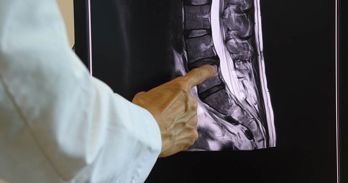
You came in with back pain, you got an MRI, and now you are holding a report with a line that reads something like: "multilevel disc desiccation with loss of disc height at L4-5 and L5-S1." You understood maybe three words. Your doctor said it was "just wear and tear" and suggested physical therapy or ibuprofen. You are still hurting and you want a clearer answer.
Over 23 years of practice in Lakewood Ranch, Dr. Banman sees this exact scenario several times a week. Patients arrive with reports in hand, confused about whether the finding means something serious, whether surgery is in their future, and whether what they are feeling is directly connected to what the radiologist wrote. The answer is rarely simple, but it is also rarely as alarming as the language sounds.
Disc desiccation is one of the most common findings on any adult lumbar or cervical MRI. It is a clue, not a verdict. Understanding what it means, and what it does not mean, is the first step toward treating the right problem. This is also closely tied to degenerative disc disease, a broader condition that disc desiccation often signals in its earlier stages.
What disc desiccation actually means
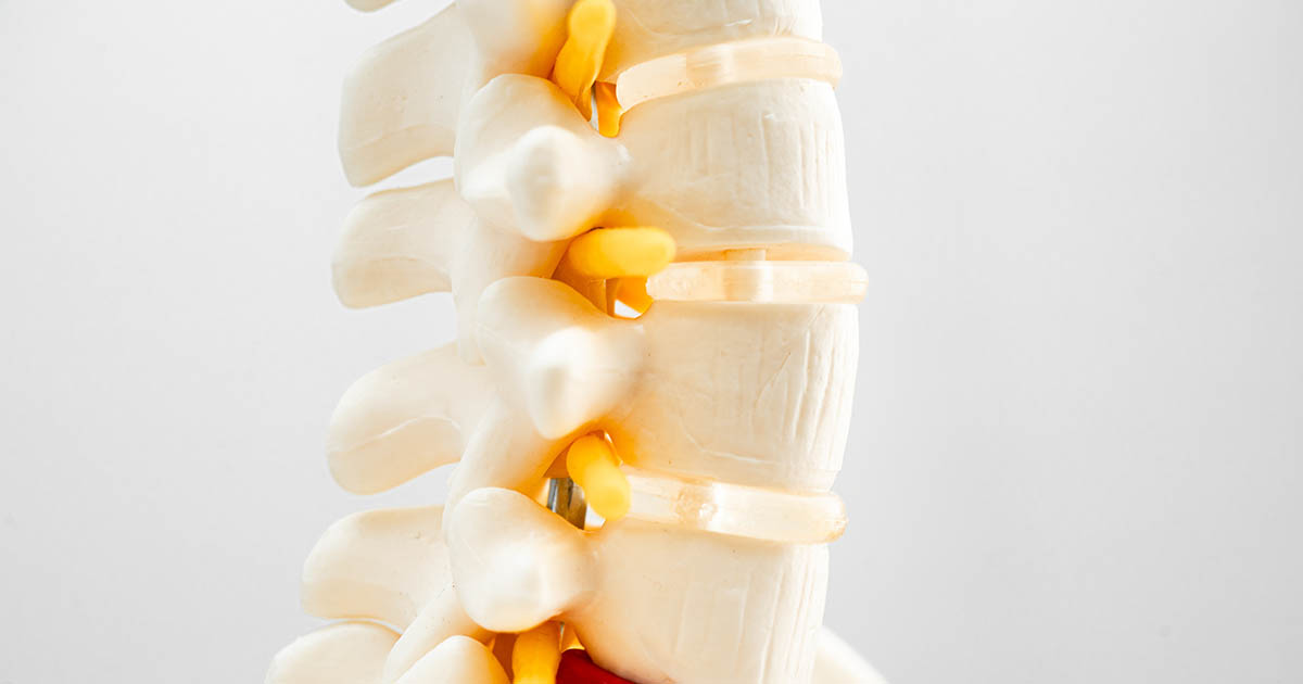
A spinal disc is essentially a sealed bag of water. The outer ring (the annulus fibrosus) is tough, fibrous, and layered like a radial tire. The inner core (the nucleus pulposus) is about 80 percent water when you are young. That water content is what gives the disc its shock-absorbing ability. Compress a well-hydrated disc and it springs back. Compress a dry one and it does not.
Desiccation is the technical term for dehydration of the nucleus pulposus. As the water content drops, the disc loses its ability to distribute load evenly across the vertebral endplates above and below it. The disc flattens. The space between vertebrae narrows. The MRI picks this up because water appears bright white on T2-weighted sequences. A hydrated disc glows. A desiccated disc looks dark gray or black. Radiologists call this the "black disc sign."
The change is gradual, measurable, and for most people starts in their 30s. By age 40, some degree of disc desiccation is present in roughly 60 to 80 percent of the population, even people with no back pain at all. By 60, near-universal. That does not make it irrelevant, but it puts the finding in context.
Why discs lose water in the first place
Discs do not have their own blood supply after childhood. They get nutrients and water through a process called diffusion, essentially soaking up fluid from the adjacent vertebral endplates when you move and lie down. Prolonged sitting, repetitive heavy loading, smoking, and poor hydration all impair that diffusion process.
Genetics play a larger role than most patients expect. Some families are simply prone to earlier or faster disc dehydration, independent of how they lift, sit, or exercise. A 35-year-old who has never done hard physical labor can have more advanced desiccation than a 55-year-old manual worker if the genes point that way.
Specific contributors include:
- Age: Proteoglycan content in the nucleus declines steadily after early adulthood, reducing water-binding capacity.
- Mechanical loading: Sustained compression without movement reduces nutrient delivery. This is part of why long-haul driving and desk work show up repeatedly in the histories of patients with early disc degeneration.
- Smoking: Nicotine constricts the small vessels that supply the endplates, reducing diffusion even further. Evidence on this link is consistent and fairly strong.
- Obesity: Excess load on the lumbar spine accelerates the mechanical stress on discs, particularly at L4-5 and L5-S1.
- Genetics: Twin studies show that 60 to 80 percent of disc degeneration variability is heritable.
What it looks like on your MRI report
Radiologists describe desiccation in varying language. You may see any of these on your report, all referring to the same underlying finding:
- "Disc desiccation" or "disc dehydration"
- "Loss of T2 signal" or "decreased T2 signal intensity"
- "Loss of disc height" or "disc space narrowing"
- "Degenerative disc changes" or "spondylotic changes"
- "Black disc sign" (less common in reports, more often a clinical shorthand)
When a report also notes "modic changes," that refers to changes in the vertebral endplates adjacent to a desiccated disc, a sign that the bone has begun to respond to the altered loading. Modic type 1 (edema) is associated with more active inflammation and more pain in many patients. Modic type 2 (fat signal) is a more chronic, stable finding.
Level matters. Desiccation at L4-5 and L5-S1 is extremely common and corresponds to the segments that bear the most compressive load in the lumbar spine. Finding it at multiple levels in the thoracic spine is less typical and sometimes warrants closer attention.
Does disc desiccation cause pain?
Sometimes. Not always. And the relationship is frustratingly imperfect.
Desiccated discs are not themselves highly innervated, but the outer third of the annulus fibrosus is. As a disc loses height and the annulus begins to develop micro-tears or fissures, those nerve endings become irritated. That produces discogenic pain, a dull, often axial ache that is hard to localize, frequently worsened by prolonged sitting, and relieved somewhat by movement or lying down.
A 2019 review in Spine Journal found that while disc degeneration on MRI is more common in symptomatic patients than in pain-free controls, the overlap is substantial. A significant portion of people with severe radiographic degeneration report no pain, and a significant portion of people with minimal MRI findings report significant pain. The scan alone does not determine treatment.
What converts a desiccated disc from an incidental finding into an active pain generator is usually a secondary change: the disc bulges and presses on a nerve root (producing leg symptoms in lumbar disease or arm symptoms in cervical), the disc loses so much height that the facet joints above and below are overloaded and inflamed, or the degenerative cascade progresses to spinal stenosis, where the overall canal narrows enough to compress the spinal cord or cauda equina.
When desiccation signals a more serious problem
On its own, mild disc desiccation at one or two levels in a 45-year-old is not an emergency. It becomes more clinically significant in these situations:
- Associated nerve root compression: If the desiccated disc has also bulged or herniated and is pressing on a nerve, you may feel pain, numbness, or weakness running into the leg (lumbar) or arm (cervical). This is distinct from the disc finding itself and changes the treatment approach. See our page on herniated disc treatment options for more detail on that overlap.
- Rapid loss of disc height: When height loss is significant enough to close down the foramen (the opening where the nerve exits), foraminal stenosis develops. This produces positional leg or arm pain that differs from classic sciatica.
- Multi-level severe desiccation in a younger patient: Finding severe desiccation at four or more levels in someone under 45 suggests either accelerated genetic degeneration or a history of heavy mechanical loading that deserves proactive management.
- Night pain or pain at rest: Disc-related pain typically improves with rest. Pain that is constant or worse at night may indicate an inflammatory or, rarely, a non-mechanical cause that needs further workup.
- Bowel or bladder changes: Loss of bowel or bladder control with back pain is a red flag for cauda equina syndrome, a surgical emergency. This is rare but requires immediate emergency evaluation.
What can actually help
There is no treatment that reverses disc desiccation in the sense of returning the disc to its original water content. What treatment does is address the consequences: the pain, the functional limitation, the downstream changes in facet joints and nerve roots, and the further progression of the degenerative cascade.
Conservative care is the right starting point for the large majority of patients with disc desiccation and back pain. The evidence consistently shows that most patients with disc-related back pain improve without surgery, though "improve" means different things to different people and the timeline varies considerably.
At our Lakewood Ranch clinic, the approach depends on what the exam reveals, not just what the MRI says. A physical examination can often determine whether pain is genuinely discogenic, whether a nerve root is involved, whether facet joints are contributing, and what movement patterns are loading the problem level most. That changes the treatment plan.
Approaches we use for disc desiccation and its downstream effects:
- Spinal decompression: Non-surgical traction-based therapy designed to create a negative intradiscal pressure that draws fluid back toward the nucleus and reduces pressure on adjacent nerve roots. For patients with disc height loss and discogenic pain, this is one of the more targeted options available outside of surgery. See our page on non-surgical spinal decompression in Lakewood Ranch for how we use it.
- Chiropractic adjustments: Restore motion to segments above and below the degenerated level, reducing compensatory overload on adjacent structures. Also relevant for facet joint pain that develops as discs lose height.
- Class IV laser therapy: Works on the inflammatory component, particularly useful when Modic changes or endplate inflammation is part of the picture.
- Rehabilitation and core stability: The disc does not hold the segment stable on its own; the muscles do. Patients with disc height loss often have compensatory muscle guarding and poor deep stabilizer activation. Addressing that directly reduces load on the disc.
- Lifestyle modifications: Quitting smoking has measurable effects on disc nutrition. Sustained low-impact movement (walking, swimming, cycling) maintains the diffusion mechanism that keeps disc cells alive. Prolonged static sitting without movement breaks is one of the worst things for an already desiccated disc.
How we evaluate disc desiccation at our clinic
When a patient brings in an MRI showing disc desiccation, the first thing we do is a thorough orthopedic and neurological examination. The MRI tells us the anatomy. The exam tells us the physiology: which movements reproduce or relieve the pain, whether nerve conduction is intact, whether the pain pattern matches the level identified on MRI, and whether anything else is contributing.
In many cases, the MRI finding and the pain source align. In some, they do not. Treating the MRI instead of the patient is one of the most common ways back pain care goes wrong. Over 23 years, Dr. Banman has seen patients who had severe radiographic degeneration respond very well to conservative care and patients with apparently mild findings who needed a completely different approach. The imaging is a piece of data, not a roadmap.
If you are in Lakewood Ranch, Bradenton, or Sarasota and you have an MRI report you want explained in plain English, along with an exam to correlate what you are feeling with what the scan shows, that is exactly what an initial visit is for. You will leave with a clear explanation of your MRI findings, an honest assessment of what is driving your symptoms, and a realistic plan.
For the bigger picture on how desiccation fits into the progression of disc disease, see our degenerative disc disease overview. If your MRI also showed a bulge or herniation at the same level, our page on herniated disc options covers the treatment decision in more detail.