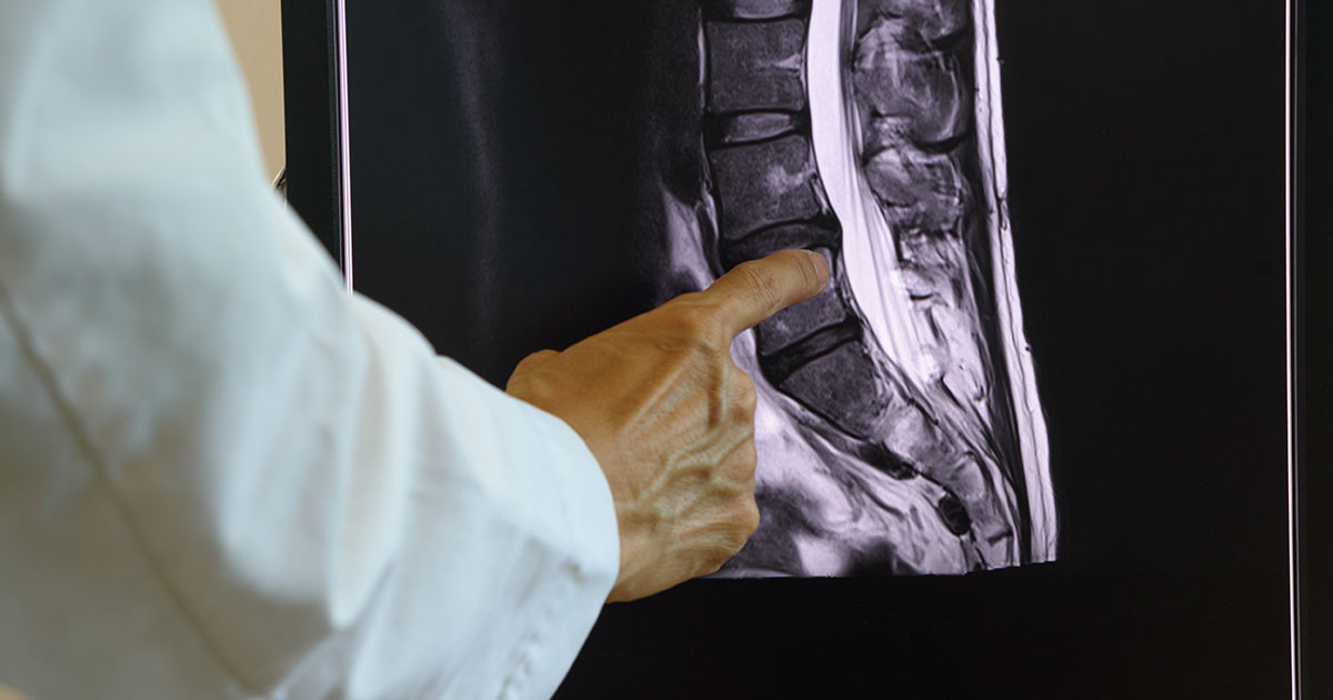
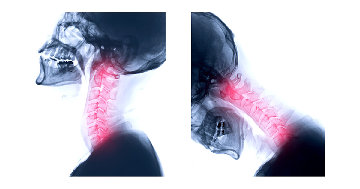
The MRI report lands in your patient portal. You scroll past the long technical section to the impression line, and somewhere in there you see it: "moderate right-sided foraminal narrowing at C5-6" or "mild foraminal stenosis bilaterally at L4-5." You search the phrase, find a wall of medical text, and close the tab more confused than when you started.
This happens constantly in our Lakewood Ranch practice. Foraminal stenosis is one of the most frequently reported MRI findings in adults over 40, and it is also one of the most misunderstood. Most patients either catastrophize it (thinking they need surgery immediately) or dismiss it (because the doctor said "degenerative changes, just normal aging"). Neither response is quite right. Understanding what foraminal stenosis actually is will help you ask better questions, make a more informed decision, and potentially avoid a lot of unnecessary treatment. For a broader overview of how spinal narrowing affects the nervous system, our spinal stenosis page covers the central canal side of that same picture.
What the Foramen Actually Is
Your spine is a column of 24 vertebrae stacked from your tailbone to the base of your skull. Between each pair of vertebrae sits a disc (the shock absorber) and, on each side of the joint, a small bony opening called the intervertebral foramen. The plural is foramina, which is why you will sometimes see "neuroforaminal stenosis" or just "foraminal narrowing" used interchangeably on reports.
These openings are the exits. Every nerve root that leaves your spinal cord on its way to your arm, leg, or torso travels through one of these foramina. The foramen is roughly kidney-shaped, about the size of your thumbnail, and it has to fit the nerve root plus blood vessels plus connective tissue, all snugly but without compression.
When the foramen narrows, the nerve root has less room. Whether that matters clinically depends on how much narrowing there is, which direction the nerve is being squeezed, and how tolerant that particular nerve happens to be.
How Foraminal Narrowing Happens
The most common causes are degenerative: they build up slowly over years. The four main contributors are:
- Disc height loss. As discs dehydrate with age (or after injury), the disc flattens. A flatter disc means the foramen gets shorter top to bottom. The nerve that used to have generous clearance now sits closer to the bone above and below it.
- Bone spurs (osteophytes). When a joint experiences repeated stress, the body lays down extra bone at the margins as a stabilizing response. These bony projections can grow directly into the foraminal space.
- Ligament thickening. The ligamentum flavum, which lines the back wall of the spinal canal, can thicken over time. If it bulges into the foramen, it takes up real estate the nerve needs.
- Disc bulge or herniation. A bulging or herniated disc that migrates toward the foramen (rather than toward the central canal) is called a "far lateral" or "foraminal" herniation. This tends to be more painful than a central herniation of the same size, because the nerve has nowhere to go.
In most cases, the finding on your MRI represents a combination of these changes over a long period. An acute traumatic event, such as a fall or a car accident, can accelerate these changes or trigger a herniation that tips a borderline foramen into symptomatic territory.
Cervical vs Lumbar Foraminal Stenosis: Different Symptoms, Same Mechanism
Where the stenosis sits in your spine determines where you feel it.
Cervical foraminal stenosis (neck) compresses nerve roots that travel into the shoulder, arm, forearm, and hand. Each level has a predictable distribution:
- C5: outer shoulder, upper arm weakness
- C6: thumb and index finger, often with biceps weakness
- C7: middle finger, triceps weakness (the most commonly affected level)
- C8: ring and little finger, hand grip weakness
Symptoms include sharp or burning pain that radiates from the neck into the arm, numbness, tingling, or weakness in the hand. If the radiating pain follows a dermatomal pattern (meaning it tracks one of these specific territories), that is a significant diagnostic clue. Our neck pain and headaches page explains how cervical nerve root compression overlaps with other causes of upper-body pain.
Lumbar foraminal stenosis (lower back) compresses the nerve roots traveling into the buttock, thigh, calf, and foot. The clinical picture looks very similar to classic sciatica. The difference is that central canal stenosis tends to cause bilateral leg heaviness and cramping with walking (neurogenic claudication), while foraminal stenosis more often produces a sharp, one-sided nerve pain that radiates down the leg following a specific nerve root pattern. Both can coexist.
The relevant lumbar levels: L4 compresses to the front of the thigh and inner calf; L5 to the outer calf and top of the foot (the most commonly affected lumbar level); S1 to the outer foot and heel, with possible weakness pushing off when walking. If you are dealing with persistent lower back pain alongside radiating leg symptoms, the distinction between foraminal and central involvement matters for how treatment is planned.
Why Your MRI Finding and Your Symptoms Often Don't Match
This is the part most patients never hear, and it matters.
Multiple large studies have imaged people with no spine symptoms whatsoever and found foraminal stenosis in a substantial percentage. One widely cited NEJM study found disc abnormalities (including foraminal narrowing) in more than half of asymptomatic adults. The rate increases with age. By your 60s, some degree of foraminal narrowing on MRI is almost expected as a normal aging finding.
The MRI shows anatomy. It does not show pain. A finding is only meaningful if it corresponds to a symptom pattern that makes clinical sense. Finding foraminal narrowing at C6-7 in a patient with C7 dermatomal pain in the right hand is significant. Finding the same narrowing in a patient whose only complaint is general neck stiffness is much less so.
That is why a good clinical examination is not optional. We look at which nerve roots, if any, show signs of irritation (reduced reflexes, dermatomal weakness, reproducible pain with specific movements). When the exam findings and the MRI findings agree, the picture sharpens. When they do not agree, the MRI finding may be real but clinically irrelevant, and chasing it with aggressive treatment often leads to worse outcomes.
This is also why a pinched nerve evaluation at our clinic always starts with the physical exam, not the imaging.
Non-Surgical Options That Produce Real Results
For the vast majority of patients with foraminal stenosis, surgery is not the first step, even when symptoms are significant. The evidence for conservative management is strong, and most patients who do eventually need surgery would not have known that without first trying non-surgical treatment for 6 to 12 weeks.
The options with meaningful clinical support:
Chiropractic adjustments. Specific spinal manipulation can reduce mechanical irritation at the affected segment, improve joint mobility, and decrease the inflammatory load on the nerve root. For foraminal stenosis specifically, the goal is not to "open" the foramen (adjustments do not permanently change bony anatomy) but to reduce the muscle guarding, joint restriction, and positional compression that amplify symptoms. In our practice, 23+ years of working with lumbar and cervical foraminal stenosis have shaped how we select technique and force levels for each patient.
Non-surgical spinal decompression. Motorized traction therapy creates a sustained negative intradiscal pressure that can reduce disc bulge into the foramen, take compressive load off the nerve root, and improve the disc's ability to rehydrate. It is not appropriate for everyone (certain fractures, severe instability, and active cancer are contraindications), but for disc-related foraminal stenosis it has a reasonable evidence base. We use it regularly. Our spinal decompression page covers how the treatment works and who tends to benefit.
Targeted exercise and postural correction. Extension-based exercises work well for some lumbar foraminal stenosis patients because extension opens the foramen slightly. Flexion-based work is often better for others. Getting the direction wrong can make symptoms worse, which is why cookie-cutter exercise sheets are not the starting point. A movement assessment first, then a tailored program.
Class IV laser therapy. Cold and Class IV laser therapy can reduce nerve inflammation and facilitate tissue healing around the compressed root. We use this as an adjunct, not a standalone, but it can meaningfully accelerate symptom resolution in the early weeks of treatment.
Activity modification (short-term). Identifying and removing the specific movements or postures that load the affected foramen gives the nerve root a chance to calm down. For many patients this is as simple as changing how they sit at a desk or how they sleep. The modification is temporary, with the goal of restoring full movement capacity as symptoms settle.
When Surgery Actually Makes Sense
Surgery is not the enemy. For the right presentation, it is often the fastest path to durable relief. The conditions that point toward surgical evaluation rather than continued conservative care:
- Progressive neurological deficit: muscle weakness is getting measurably worse over weeks, not just pain that varies day to day.
- Bowel or bladder dysfunction: sudden inability to control bowel or bladder is a red flag requiring emergency evaluation, not a scheduled appointment.
- Myelopathy signs: if the stenosis affects the spinal cord (not just the nerve root), symptoms include difficulty with fine motor tasks, gait changes, and hand clumsiness. This is a different and more urgent situation.
- Failure of 6-12 weeks of active, well-directed conservative treatment with no meaningful improvement in function.
If surgery is discussed, the common procedures for foraminal stenosis are foraminotomy (surgical enlargement of the foramen) and, when a disc herniation is the driver, microdiscectomy. These are generally well-tolerated with good outcomes when the diagnosis is accurate. The problem arises when surgery is performed based on imaging alone, without a clear clinical correlation. The imaging finding stays after the surgery; if it was not the pain generator, neither does the pain resolve.
What to Do After Getting This Finding on Your MRI
Here is a practical sequence:
- Get a clinical examination that maps to the MRI level. Someone should check your reflexes, test muscle groups that correspond to the affected nerve root, and reproduce your symptom pattern with specific movements. If the MRI level matches the clinical findings, you have a real problem to address. If it does not, the finding may be a bystander.
- Start conservative treatment before committing to anything permanent. Most insurers require documented conservative care before approving surgery anyway. More importantly, most patients do not need surgery after completing a proper trial of conservative care.
- Track function, not just pain. Can you carry groceries, sit through a meeting, sleep a full night? Progress in those measures matters more than whether the pain is a 4 or a 5 on any given day.
- Re-evaluate if things are not moving by week 6-8. A direction change at 6 weeks is reasonable; a direction change at 6 days is not. Give the conservative plan enough time to show results, but not so much time that a progressive deficit is allowed to continue unchecked.
If you have an MRI report in hand and want a second opinion on what it means clinically, that is something we do routinely in Lakewood Ranch. Dr. Banman has reviewed hundreds of spine MRI reports alongside physical exams over his 23+ years in practice. The reading takes about 20 minutes and generally tells you clearly which direction makes sense.