You just got your MRI results back. The report says something like "disc herniation at L4-L5 with moderate left foraminal narrowing" or "L5-S1 disc protrusion with S1 nerve root compression." You probably searched for what that means and found a wall of medical jargon. Let me break it down in plain language.
In 23-plus years of treating spinal disc injuries at Spine and Wellness Center in Lakewood Ranch, the two levels I see most are L4-L5 and L5-S1. Together they account for roughly 90 percent of lumbar disc herniations. They are at the bottom of your lumbar spine, they carry the most mechanical load, and when they herniate, they press on different nerve roots that travel down your leg in specific, mappable patterns.
Knowing which level is affected changes the conversation about treatment. It tells us which nerve to decompress, which movement patterns are putting it under stress, and what the realistic recovery window looks like. If you have been diagnosed with a disc problem and your leg pain or numbness does not quite make sense yet, this page is for you. For a broader look at herniated disc treatment options at our clinic, start there.
Why the Disc Level on Your MRI Report Matters
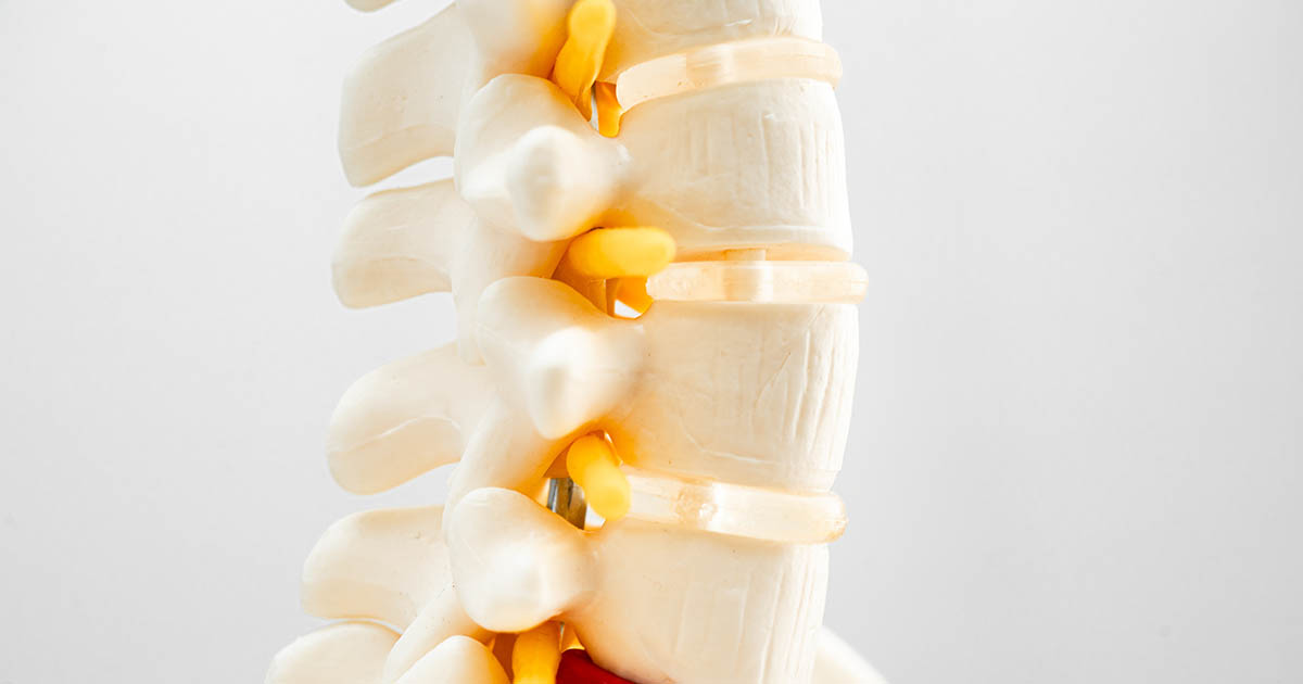
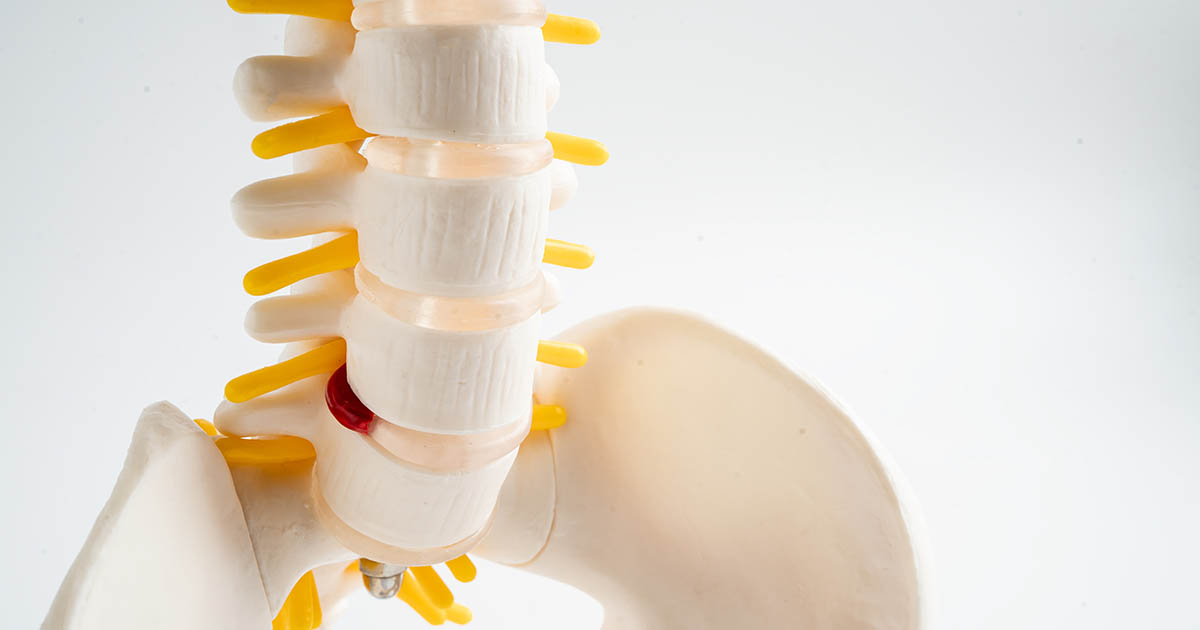
Your lumbar spine has five vertebrae, labeled L1 through L5 from top to bottom. Below L5 sits the sacrum, which begins with S1. Between each pair of vertebrae sits a disc: a fibrous outer ring (annulus fibrosus) with a gel-like center (nucleus pulposus). That disc acts as a shock absorber and spacer.
When the outer ring weakens, the inner material can push outward. When that material pushes toward the spinal canal or the nerve exit points (foramina) on the side, it contacts a nerve root. That contact is what causes the classic symptoms: pain shooting down the leg, numbness in a specific zone, or weakness in a particular muscle group.
Here is the key: each disc level sits next to a specific nerve root. A herniation at L4-L5 most commonly presses on the L5 nerve root. A herniation at L5-S1 most commonly presses on the S1 nerve root. Those two nerves travel to entirely different parts of your foot and leg. Mixing them up means chasing the wrong symptom pattern.
The level on your MRI report is not just anatomy trivia. It predicts exactly where your numbness should be, which movements should aggravate it, and which muscles we check first during the exam.
The L4-L5 Level: L5 Nerve Root
L4-L5 is the second-to-last disc in your lumbar spine. It is one of the most mobile segments in the lower back, which is partly why it is so frequently injured. A herniation here, whether central, paracentral, or foraminal, typically contacts the L5 nerve root.
The L5 nerve root is the one that travels along the outer shin, over the top of the foot, and into the big toe and the two toes beside it. When it is compressed, the symptoms follow that pathway.
What you typically feel with an L4-L5 herniation:
- Pain starting in the low back or buttock, tracking down the outer thigh and outer shin to the top of the foot
- Numbness or tingling at the top of the foot, around the big toe and second toe
- Weakness lifting the foot upward (dorsiflexion): difficulty clearing the floor when walking, which in severe cases becomes foot drop
- Weakness extending the big toe: one early test is trying to push your big toe upward against resistance
- Pain that worsens when sitting for long periods or with lumbar flexion (bending forward)
One thing that surprises many patients: the L5 nerve root does not have a strong reflex associated with it. Your knee jerk (patellar reflex) belongs to L4, and your ankle jerk (Achilles reflex) belongs to S1. So a provider who only checks those reflexes may not catch early L5 compression. The more reliable tests are strength and sensation in the specific L5 distribution.
If your sciatica runs down the outer leg and over the top of your foot, L4-L5 is the most likely culprit.
The L5-S1 Level: S1 Nerve Root
L5-S1 is the very bottom of your lumbar spine, the junction between your last lumbar vertebra and the sacrum. It handles even more compressive load than L4-L5, especially during prolonged sitting. When the L5-S1 disc herniates, it most commonly compresses the S1 nerve root.
The S1 nerve root travels down the back of the thigh, through the calf, and into the heel and outer edge of the foot, including the little toe. The distribution is distinctly different from L5.
What you typically feel with an L5-S1 herniation:
- Pain tracking down the center or back of the thigh, through the calf, and into the heel or the sole and outer foot
- Numbness or tingling along the outer ankle, the heel, or the bottom of the foot
- Weakness pushing off the ball of the foot (plantarflexion): difficulty rising up on tiptoe, or a feeling that the calf is not generating its normal power
- A reduced or absent ankle jerk (Achilles reflex): one of the most clinically useful signs of S1 compression, because this reflex is easy to test and reliable
- Pain that often increases with prolonged standing or walking and eases slightly with lumbar extension
The calf muscle is largely S1 territory. Many patients with L5-S1 herniations describe calf heaviness, cramping, or a sense that the leg just does not have the same push it used to. If you have been struggling with what feels like calf pain but no obvious muscle injury, the S1 nerve root is worth investigating.
Comparing L4-L5 and L5-S1: A Practical Guide
Here is a side-by-side look at the key differences. Keep in mind that real presentations overlap, and many patients have herniations at both levels simultaneously. This is a guide to the typical pattern, not a diagnostic rule.
- Pain location: L4-L5 (L5 nerve) sends pain down the outer shin and top of the foot. L5-S1 (S1 nerve) sends pain down the back of the leg and into the heel or sole.
- Numbness zone: L5 affects the top of the foot and the big toe area. S1 affects the heel, outer ankle, and little toe side of the foot.
- Muscle weakness: L5 compression shows up as difficulty lifting the foot or big toe. S1 compression shows up as reduced push-off strength and calf weakness.
- Reflex change: L5 compression usually does not change a standard reflex. S1 compression often reduces or eliminates the ankle jerk.
- Aggravating position: L4-L5 pain often worsens with flexion (sitting, bending). L5-S1 pain often worsens with extension (standing, walking).
A careful clinical exam plus the MRI together usually make the picture clear. If the exam findings match the MRI level, we have a high degree of confidence in the diagnosis and can target care accordingly.
What Your MRI Report Does Not Tell You
Here is something most patients do not know until after their first consultation: a herniated disc on MRI does not automatically mean surgery, and the size of the herniation on film does not always predict how much pain you are in.
Studies have found that a large percentage of adults with no symptoms at all have disc herniations on MRI. Conversely, some patients with severe symptoms have what looks like a relatively modest herniation on imaging. What actually determines your symptoms is whether the herniated material is actively compressing a nerve, how inflamed that nerve is, and how your spine is loaded and moving.
The MRI also does not tell you how the disc is being loaded. Two patients can have the same L4-L5 disc herniation on film and have completely different outcomes based on their movement patterns, core stability, and how they sit, stand, and sleep. That is why an individualized examination for lower back pain matters as much as the imaging itself.
The disc level tells us where to look. The exam tells us what to do about it.
Nonsurgical Care Options for L4-L5 and L5-S1 Herniations
The majority of lumbar disc herniations, including those at L4-L5 and L5-S1, respond to nonsurgical care. Surgery is typically reserved for cases with progressive neurological loss (muscle weakness getting worse over days to weeks), loss of bladder or bowel control, or months of failed conservative care. Outside those scenarios, the evidence supports trying structured conservative care first.
At Spine and Wellness Center, the approach we use most often for disc herniations at these two levels includes:
- Spinal decompression therapy: A motorized traction table that gently creates negative pressure inside the disc, drawing the herniated material back toward center and reducing nerve compression. We typically pair it with positioning that takes the specific affected disc level off load. For a detailed look at how this works, see our page on spinal decompression in Lakewood Ranch.
- Chiropractic adjustments: Restoring proper motion to the segments above and below the herniated level reduces the mechanical stress the injured disc is absorbing. This is not about "cracking" the herniated disc; it is about improving the mechanics of the whole lumbar region.
- Class IV laser therapy: Applied directly over the affected nerve root and disc, high-intensity laser reduces local inflammation and accelerates tissue healing. Many patients notice a meaningful reduction in nerve pain within the first several sessions.
- Rehabilitation exercises: Once the acute nerve pain settles, specific loading exercises (the exact type depends on whether the herniation is better with flexion or extension) help rehydrate the disc and build the stability to prevent recurrence.
The sequence and emphasis differ between L4-L5 and L5-S1 cases. An L5-S1 patient with calf weakness and a reduced ankle reflex gets a different exercise prescription than an L4-L5 patient with foot drop risk. That specificity is one reason the disc level matters clinically, not just on the report.
Red Flags That Need Urgent Attention
Most lumbar disc herniations are painful and uncomfortable but not dangerous. The exceptions involve the cauda equina, a bundle of nerve roots in the lower spinal canal that controls bladder, bowel, and sexual function.
If you have any of the following, go to the emergency room or call your physician the same day. These are not situations for conservative care first:
- Loss of bladder or bowel control (or difficulty sensing when you need to go)
- Numbness or tingling in the groin, inner thighs, or perineal area (saddle anesthesia)
- Rapid progression of leg weakness over 24 to 48 hours
- Foot drop that appeared suddenly rather than building gradually
Everything else, including pain that makes you miserable but leaves neurological function intact, is a candidate for conservative care with appropriate monitoring. We always re-examine at regular intervals, so if something is not improving or is worsening, we know quickly and can refer for surgical evaluation.
What to Do If You Have an L4-L5 or L5-S1 Herniation
Get a clear picture of your neurological status before you commit to any treatment path. That means an exam that tests strength, sensation, and reflexes in the specific distribution of the nerve your MRI implicates, not just a general "check your reflexes" evaluation.
In our office in Lakewood Ranch, the initial disc evaluation includes a posture and range-of-motion assessment, orthopedic disc tests, and a nerve tension test series (straight-leg raise, Bragard's, seated slump). We review your imaging alongside the physical findings so that the MRI findings and the exam findings either confirm each other or raise the right questions. Many patients tell us it is the first time anyone has explained the connection between what they feel and what the MRI shows.
If you have already been told "your only option is surgery" but you have not yet tried structured conservative care, it is worth getting a second opinion. The research on lumbar disc herniations at L4-L5 and L5-S1 consistently shows that the majority of patients who complete a proper course of decompression-based care avoid surgery entirely. In our experience with patients across the Lakewood Ranch, Bradenton, and Sarasota areas, that outcome holds more often than not.