You have a stack of papers from the imaging center. The first line says: "L4-5 posterior disc herniation with moderate right foraminal narrowing and mild facet arthrosis. L3-4 diffuse annular bulge without significant canal compromise. Mild degenerative endplate changes at L2-3." You read it twice. You understand nothing. Your doctor's office said someone would call in three to five business days.
That is a genuinely frustrating situation. The MRI is a picture of your body, your pain, your problem. You deserve to understand what it says before anyone starts explaining options. In 23-plus years of practice here in Lakewood Ranch, I have seen this same scenario dozens of times: someone arrives with a report they received weeks ago, having quietly worried about the word "herniation" or "severe" without knowing what those words mean in context. Our back pain overview covers the most common structural drivers; this post gives you the vocabulary to understand what imaging actually found.
A quick map of what the MRI is actually showing
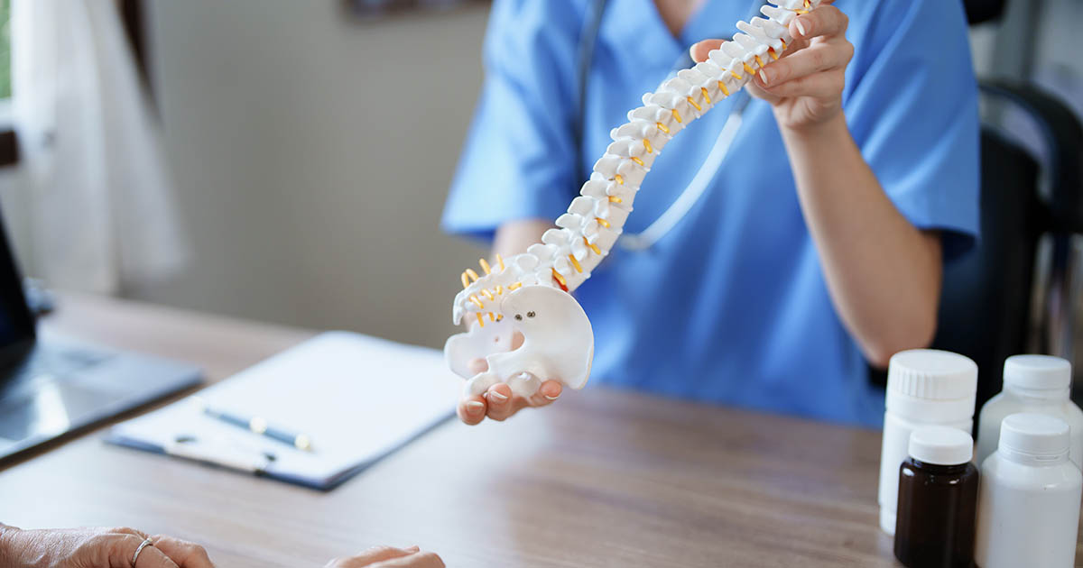
Your spine has 33 vertebrae, grouped and named by region. The cervical spine (C1 through C7) is your neck. The thoracic spine (T1 through T12) is your mid and upper back, where the ribs attach. The lumbar spine (L1 through L5) is your lower back, where most disc problems occur. Below that, the sacral and coccygeal vertebrae are fused into fixed structures.
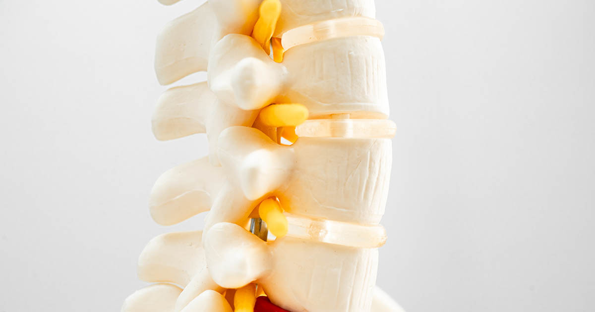
Between each pair of vertebrae sits an intervertebral disc: a tough outer ring called the annulus fibrosus wrapped around a softer, gel-like core called the nucleus pulposus. The disc acts as a shock absorber and maintains the space between vertebrae, which matters because that space is where nerve roots exit.
Running through the bony vertebrae is the spinal canal, a tunnel that protects the spinal cord down to roughly L1-L2 and the cauda equina (a bundle of individual nerve roots) below that. At each level, pairs of nerve roots branch off through small side openings called foramina. When something narrows those openings, symptoms can radiate into an arm or leg.
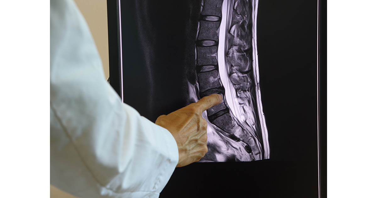
MRI shows soft tissue in detail: discs, ligaments, the spinal cord, nerve roots, surrounding muscles. A plain X-ray shows bone clearly but misses most of the structures that actually generate pain. That is why imaging for back and leg symptoms typically starts with MRI rather than X-ray alone.
What those disc findings actually mean
Most of the confusing language in a radiology report describes what is happening to the discs. Here is the spectrum from least to most structurally significant:
Disc desiccation. The disc has lost hydration. A healthy disc in a young adult is roughly 80% water, which gives it height and shock-absorbing capacity. With age and repetitive load, discs dry out and lose some height. On T2-weighted MRI sequences, desiccated discs appear darker rather than the bright white of a well-hydrated disc. Desiccation alone rarely causes severe pain; it signals that the disc has less reserve, not that it is acutely injured.
Annular bulge or diffuse bulge. The outer ring of the disc has expanded outward uniformly around its circumference. Think of squeezing a burger patty until the edges push out in all directions. A diffuse bulge typically does not directly compress a specific nerve root; it can cause local disc inflammation and dull, central back pain without the radiating pattern of nerve involvement. "Mild disc bulge" is an extremely common finding in adults over 40, present in many people with no pain at all.
Focal protrusion. A localized bulge affecting less than half the disc's circumference. The annulus is still intact, but the disc material pushes in one direction. Whether this matters depends on where it pushes: a central protrusion is less likely to contact a nerve root than a far lateral one aimed directly at the foramen.
Herniation. The inner nucleus has pushed through a tear or weak point in the outer annulus. This is what people usually mean by "slipped disc," though nothing actually slips. The extruded material can press on the spinal cord or, more commonly, on a nerve root as it exits. When a herniated disc contacts a nerve root, it creates both mechanical compression and chemical irritation from inflammatory compounds released by the disc material. That combination is what drives the sharp, radiating leg or arm pain many patients describe. For what a herniation means for treatment, see our herniated disc page.
Extrusion. The herniated material has gone beyond the disc space and extends up or down behind the vertebra, but remains connected to the disc. Larger than a typical herniation and more likely to significantly contact neural structures.
Sequestration. A fragment has broken completely off the disc and migrated freely in the spinal canal. Less common. The free fragment may move away from the nerve root on its own over time, which is one reason many disc problems improve without surgery, but the acute presentation can be quite severe.
The word "herniation" spans a large range. A report that says "small central disc herniation" is describing something quite different from "large right paracentral disc extrusion with significant cord compression." The severity language and the location within the disc (central, paracentral, foraminal, extraforaminal) together tell the clinical story.
Foraminal narrowing and central stenosis: when does channel size matter?
Foraminal stenosis (also called foraminal narrowing or neural foraminal compromise) means the side channel where a nerve root exits has narrowed. This can come from a disc bulge or herniation pushing into the foramen, from bone spurs growing there, or from ligament thickening. The nerve root can be pinched, causing radiating pain, numbness, or weakness in the arm or leg served by that root. "Moderate right L4-5 foraminal stenosis" means the right-side exit hole at the junction of the fourth and fifth lumbar vertebrae is narrowed to a moderate degree.
Central canal stenosis means the main spinal canal has narrowed, putting pressure on the cord (in the neck or mid back) or on the cauda equina (in the lower back). When central stenosis affects the lumbar spine, the classic symptom pattern is neurogenic claudication: aching, heaviness, or weakness in both legs that worsens with walking or standing and eases when you sit or lean forward on a shopping cart. That forward-flexed position opens the canal slightly and relieves pressure. See our spinal stenosis page for how this pattern differs from disc herniation pain and what conservative care looks like.
The words "mild," "moderate," and "severe" are relative assessments, not absolute measurements. Two radiologists reviewing the same scan sometimes use different grades. More clinically useful is whether the imaging findings correspond to your actual symptoms. A "severe" foraminal narrowing on the left side may be irrelevant if all of your symptoms are on the right, and a "mild" disc herniation directly contacting a nerve root may explain significant pain.
Degenerative changes: what the vocabulary actually describes
Degenerative disc disease (DDD) sounds alarming but is largely an age-related process. It describes a disc that has lost height, hydration, and some structural integrity. Most adults over 50 have at least mild DDD in their lumbar spine; many have no pain from it whatsoever.
Osteophytes and bone spurs are bony outgrowths that form along vertebral edges in response to instability, altered load distribution, or chronic wear. They are the body's attempt to stabilize a joint it perceives as too mobile. Small spurs cause no problem; spurs that grow into the foramen or central canal narrow the available space for neural structures and can contribute to stenosis symptoms.
Modic changes are signal changes in the endplates: the bone surfaces that touch the disc. Type 1 Modic changes indicate active marrow edema and inflammation and do correlate reasonably well with pain. Type 2 indicates fatty replacement (more stable, less acutely painful). Type 3 indicates bone sclerosis. Seeing "Modic type 1 changes at L5-S1" on a report is one of the findings that does tend to have a clinical meaning worth discussing with your provider.
Ligamentum flavum hypertrophy refers to thickening of the yellow ligament that lines the back wall of the spinal canal. It is a common contributor to central stenosis in older adults because the ligament buckles inward as the disc loses height, reducing the canal diameter from the posterior side.
When the MRI says one thing and your body says another
A report that says "severe degenerative changes" does not mean your spine is beyond help. It means the disc and surrounding structures have changed over time. What matters more is what is actually generating your current pain, which is determined by a clinical examination, not by imaging alone. The scan tells us where to look; the exam tells us what to do.
MRI findings and pain levels do not always match, and this is worth knowing before your follow-up appointment. Studies going back decades have found significant disc abnormalities in people with no pain at all. In one frequently cited study in the New England Journal of Medicine, 64% of asymptomatic adults had disc abnormalities on MRI. That number climbs with age. The presence of a finding on imaging does not automatically mean it is the source of your pain.
Conversely, some patients have severe pain from modest imaging findings because the mechanism is partly chemical rather than purely mechanical. When disc material herniates, it releases inflammatory cytokines that irritate a nerve root even without significant physical compression. This is one reason people can have debilitating sciatica from a relatively small herniation.
For patients dealing with sciatica, the clinical exam, symptom distribution, and response to position changes often tell us as much as the scan does about which level is responsible and whether a nerve root is involved.
Red flags in a radiology report: a short list
Most findings can wait for a standard follow-up. A few warrant faster attention:
- Cord compression with signal change: If the spinal cord itself shows abnormal T2 signal (not just contact), this can indicate myelopathy, a condition where cord function is compromised. Cervical myelopathy in particular can progress and warrants a neurosurgical or orthopedic spine consult sooner rather than later.
- Cauda equina compression: A large central herniation compressing the full bundle of nerve roots at the lower lumbar level. If this comes with new bowel or bladder dysfunction, saddle-area numbness, or sudden profound leg weakness, that is a potential surgical emergency, not a condition to manage conservatively while waiting.
- "Cannot exclude" or "further workup recommended" language: Radiologists use this phrasing when an incidental finding warrants additional imaging or clinical follow-up but is not definitively abnormal on the current scan. Do not ignore these notations.
These are not common findings. The vast majority of lumbar MRI reports describe disc and degenerative changes that are addressed with conservative care and rarely require surgery.
How we use your MRI at the clinic
When a patient brings an MRI to our Lakewood Ranch office, we review the report alongside the clinical picture: where you hurt, what makes it better or worse, whether the pain radiates into an arm or leg, what the hands-on examination shows, and what your daily function looks like. Imaging guides several key decisions:
- Which spinal levels are most involved structurally
- Whether a disc herniation is contacting a specific nerve root, and which one
- Whether the spine is stable enough for specific manual techniques
- Whether the findings warrant referral to a specialist alongside conservative care
For disc herniations that are contacting a nerve root, non-surgical spinal decompression in Lakewood Ranch creates a sustained, computer-guided traction force that reduces intradiscal pressure and encourages retraction of herniated material. The DOC-20 table we use allows us to target specific lumbar levels rather than applying a generalized stretch. We typically combine decompression with Class IV laser therapy to address the inflammatory component and with progressive rehabilitation to restore the strength and movement patterns that protect the disc long-term.
We are also careful about what MRI cannot tell us: it cannot measure how much pain you are in, how much function you have lost, or how a finding is affecting your specific life demands. That context comes only from talking with you.
If you have an MRI report that was handed to you with minimal explanation, or if you have had back or leg pain for more than a few weeks and imaging has not yet been ordered, a consultation is a reasonable next step. Bring the images on CD or digital format if you have them, not just the paper report.