Leg Pain When Walking That Goes Away With Rest: Stenosis or Circulation?


The pattern is unmistakable. You walk a block, maybe a quarter mile, and your legs start burning, cramping, or feeling impossibly heavy. You stop for a minute, the pain fades, and you walk again. It comes back at roughly the same point every time.
This symptom has a clinical name: claudication. It is produced by two completely different conditions, and the treatment for one does essentially nothing for the other. If a doctor has told you this is "just circulation," that may be exactly right. Or it may be a problem in your lower spine pressing on the nerves that supply your legs. Getting the distinction right is not a minor detail. It determines whether you need a cardiologist, a chiropractor, or eventually a surgeon.
Two Systems That Produce the Same Symptom
Claudication (from the Latin word for limping) describes leg pain, cramping, or weakness that appears with walking and clears with rest. Two mechanisms create this cycle:
- Vascular claudication comes from peripheral arterial disease (PAD). Plaque has narrowed the arteries supplying the leg muscles. During walking, working muscles need more oxygen than narrowed arteries can deliver. Pain arrives. The moment you stop, oxygen demand drops, the supply catches up, and the pain clears.
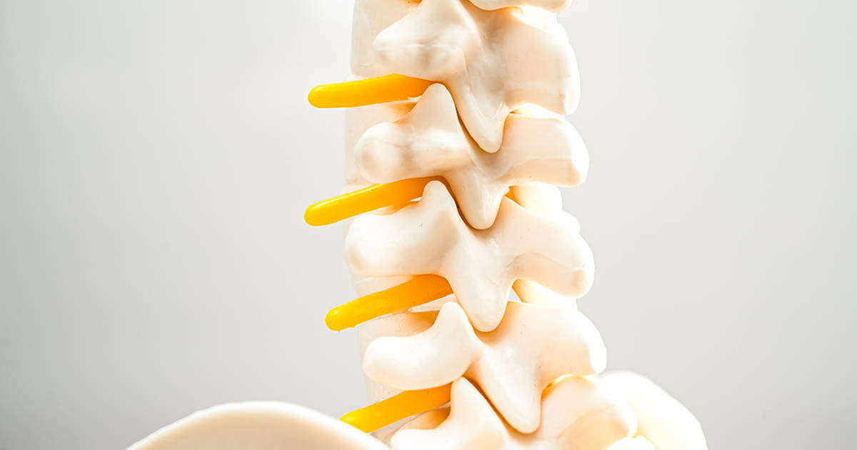
- Neurogenic claudication comes from lumbar spinal stenosis. The spinal canal has narrowed (from bone spurs, thickened ligaments, or disc bulging), which compresses the nerve roots that control sensation and strength in the legs. Walking loads the lumbar spine, temporarily worsening the compression. Rest reduces it.
On paper the two sound nearly identical. In practice, a handful of clinical features separate them almost every time.
The Single Most Useful Question
Ask yourself: does the leg pain clear up when you simply stop walking and stand in place?
If standing still is enough, the likely cause is vascular. The muscles stop working, oxygen demand drops to baseline, and the cramping resolves within two to five minutes regardless of your posture.
If you have to sit down or lean forward before the pain goes away, the cause is more likely neurogenic. Forward flexion opens the lumbar spinal canal. Standing upright does not. The nerve roots stay compressed until you change spinal position.
This one difference explains a lot of otherwise puzzling behavior. People with lumbar stenosis often push a grocery cart leaning far over the handle. They find walking uphill easier than flat ground. They can ride a stationary bike for long stretches, because the seated, slightly hunched posture keeps the spine in flexion. People with vascular claudication show none of these postural preferences. Their constraint is artery caliber, not spinal geometry.
Vascular Claudication: What to Know
PAD is an atherosclerotic disease. The same process that narrows coronary arteries can narrow the iliac, femoral, and popliteal arteries supplying the legs. Risk factors overlap almost completely with heart disease:
- Smoking (the strongest single predictor)
- Diabetes mellitus
- High blood pressure
- High LDL cholesterol
- Age over 50, especially in men
The cramping is usually calf-dominant, reflecting the calf's high oxygen demand during walking, though the thigh or buttock can be involved depending on which artery is narrowed. Onset distance tends to be predictable: the same person cramps at the same point on the same route under similar conditions. There is no back pain, no tingling or numbness, and no change when stooping or bending. The legs ache and stop working until blood flow catches up.
Diagnosis uses the ankle-brachial index (ABI), a non-invasive Doppler measurement that compares blood pressure at the ankle to blood pressure in the arm. A ratio below 0.9 indicates arterial disease. Duplex ultrasound, CT angiography, or MR angiography can pinpoint where the narrowing is. Management is cardiovascular: supervised walking exercise (counterintuitive but robustly effective), smoking cessation, blood sugar and blood pressure control, statins, and antiplatelet agents. Severe cases may require angioplasty, stenting, or bypass surgery.
Neurogenic Claudication: What to Know
Lumbar spinal stenosis is one of the most common spinal diagnoses in adults over 60, though it can develop earlier. The central canal, the lateral recesses, or the foramina through which nerve roots exit can all be narrowed by:
- Facet joint arthritis and bone spur formation
- Hypertrophy (thickening) of the ligamentum flavum
- Disc bulging or herniation
- Spondylolisthesis, where one vertebra slips forward on the next
Compression on the nerve roots increases during lumbar extension (standing upright, walking on flat ground or downhill, reaching overhead) and decreases during flexion (sitting, leaning forward, lying in the fetal position). This is why neurogenic claudication is so reliably position-dependent.
The symptom pattern tends to be broader than in vascular claudication. People describe aching, heaviness, burning, or numbness in the buttocks, thighs, calves, or feet. It is sometimes bilateral and sometimes asymmetric. Low back pain is often present and can predate the leg symptoms by years. The walking distance before symptoms appear varies with posture and terrain: longer when leaning forward, shorter when standing fully upright, longer going uphill, shorter going downhill.
Imaging confirms the diagnosis. An MRI of the lumbar spine shows the degree of canal narrowing, which levels are affected, and whether nerve roots are being compressed. X-rays with flexion-extension views show instability or spondylolisthesis that an MRI can miss.
Why These Two Conditions Get Confused
Both are more common in older adults, so they coexist in the same person more often than chance alone would predict. Someone in their 60s or 70s with a history of smoking and diabetes may have PAD and lumbar stenosis at the same time. In that case, the claudication distance is shorter than either condition alone would produce, and neither intervention fully resolves the symptoms on its own.
A few other conditions can produce a similar walking-rest-walk pattern:
- Venous claudication: from chronic venous insufficiency or prior deep-vein thrombosis. The leg feels bursting and congested rather than cramped, and elevation helps more than forward flexion.
- Hip osteoarthritis: groin and anterior thigh pain with walking, but it does not resolve fully with brief rest and is reproduced by rotating the hip at the end of range.
- Cauda equina syndrome: a rare but serious compression of the nerve bundle at the base of the spinal cord; the distinguishing feature is bladder or bowel dysfunction, which requires immediate evaluation.
Ordering the wrong test first is the most common source of delay. A person with neurogenic claudication who gets only an ABI will have a normal result and be sent home without an answer. A person with PAD who gets only a lumbar MRI may be told they have "degenerative changes" (present in more than 90 percent of adults over 60 and often entirely asymptomatic) and be treated for the wrong problem. A thorough clinical history and a targeted exam narrow the field before a single imaging study is ordered.
Conservative Care for Neurogenic Claudication
Lumbar stenosis does not inevitably require surgery. Randomized controlled trials have found that patients with moderate stenosis who follow a structured conservative care program reach similar functional outcomes to surgical patients over two to four years, though the trajectory is slower. Surgery becomes the appropriate path when conservative care has been fully pursued, or when a patient shows progressive muscle weakness or signs of bladder and bowel involvement.
Conservative options that have meaningful evidence or clinical rationale include:
- Spinal decompression therapy: Motorized traction applies a controlled distraction force to the lumbar spine, reducing intradiscal pressure and creating space within the narrowed canal. A treatment course typically involves multiple sessions over several weeks, with progress assessed at intervals.
- Chiropractic adjustments: Restore normal motion in joints that have stiffened around a stenotic segment. Restricted adjacent segments often compensate in ways that increase load at the narrowed level; restoring their mobility reduces that load.
- Flexion-distraction technique: A specific manual method that cycles the lumbar spine through repetitive flexion while applying gentle traction, directly widening the canal in a controlled and low-force way. It is well-tolerated even in older patients with significant arthritis.
- Targeted strengthening: Building the capacity of muscles that stabilize the spine in neutral or slight flexion reduces the extension load that narrows the canal during walking. Exercises are selected to avoid spinal extension during the active phase of care.
Epidural steroid injections can reduce nerve root inflammation and extend walking tolerance, sometimes substantially. Their effect is frequently temporary, so they are often used as a bridge to allow a patient to participate in conservative care rather than as a stand-alone treatment.
Starting With the Right Question
The clinical history is where differentiation begins. When does the pain come on, where does it go, how long does rest relieve it, does leaning forward help, is there back pain, does the pain change with stairs or hills? Those seven questions, answered carefully, point toward one condition or the other in most cases before any test is run.
Both conditions are manageable. Neither improves by waiting for the diagnosis to become obvious. The person who can walk only to the end of the driveway before stopping has a problem that is limiting their life today, and identifying the mechanism is what opens the door to a solution.
At Spine and Wellness Center Lakewood Ranch, we evaluate structure, movement, and nerve function together, because the same symptom can have very different origins. Knowing which one you are dealing with is not a fine point. It is the entire question.